Most patients are able to function and effectively with a maxillary complete denture. However, some patients with reasonable bearing surfaces will benefit from implant retained overdentures such as those where peripheral seal is difficult to achieve and maintain. Others will prefer an implant retained prosthesis for psychological or psychosocial reasons. In still other patients with opposing natural dentition in the mandible, implants are justified on the basis of providing additional support or resistance to the forces of chewing in order to prevent resorption of bone of the edentulous maxilla. This program of instruction provides useful insights into treatment planning, prosthesis design and the most common methods used to retain the prosthesis.
1. 6. Edentulous Maxilla Overlay Dentures John Beumer III DDS, MS Hiroaki Okabe CDTDivision of Advanced Prosthodontics, Biomaterials and Hospital Dentistry, UCLAThis program of instruction is protected by copyright ©. No portion ofthis program of instruction may be reproduced, recorded ortransferred by any means electronic, digital, photographic, mechanicaletc., or by any information storage or retrieval system, without priorpermission.
2. Patient selection and treatment planning Implant Assisted vs Implant Supported Fixed vs Removable Overlay Dentures !Fixed detachable !Implant assisted prosthesis (Implant! Fixed prosthesis overdentures supported overlay dentures with milled bars)
3. DefinitionsImplant assisted prosthesisThe forces of occlusion are sharedbetween the implants and themucoperiosteum. Alwaysremovable overlay dentures.Implant supported prosthesisAll the forces of occlusion areborne by the implants. Can beeither fixed partial dentures orremovable overlay dentures.
4. Edentulous MaxillaMost patients are best served withimplant assisted overlay dentures. Why?
5. Treatment Planning and Patient Selection Edentulous Maxilla ! Bone quantity and quality ! Resorptive patterns ! Maxillomandibular relations ! Sinus architecture ! Esthetics/lip support ! Phonetics ! Hygiene access and patient compliance ! Cost ! Predictability of surgical interventionsIn most patients careful consideration of these factors will favor implant assisted removable overlay dentures.
6. Resorptive patterns and maxillo- mandibular relationships Fixed vs Removable The normal patterns of resorption result in pseudo class III jaw relations. Overlay dentures (either implant supported or implant assisted) are preferred over fixed prostheses. Why?Removable overlay dentures with properly extended and contoureddenture flanges provide better lip support and facial contours.
7. Esthetics/Lip Support Most elderly patients require the lip support provided by the labial flange of an overlay denture particularly if they have been wearing a maxillary denture for an extended length of time.
8. Esthetics – Lip Support Fixed vs Removable ! Fixed in retrospect was a poor choice ! As the patient aged and lost tonus of the lip musculature lip contours became deficient
9. Esthetics – Lip Support Fixed vs Removable Fixed was an appropriate choice for this patient ! More favorable jaw relationship. Lip support from the prosthesis is not required to same degree as the previous patient. ! Low smile line meant the proximal spaces designed for hygiene access are not visible.
10. Esthetics – Lip Support Fixed vs RemovableLip line v If the patient presents with a high lip line they are probably best served by an overdenture
11. Phonetics – Fixed vs Removable! This design was used frequently in the 80’s. Note the spaces beneath the bridge. These spaces facilitate hygiene but during speech air escapes through these spaces adversely affecting speech articulation. Removable overlay dentures prevent this problem.
12. Fixed vs RemovableThe speech vs hygiene dilemma v If you close the spaces and provide lip support access for oral hygiene is compromised v If you provide hygiene access, you compromised lip support and speech articulaion
13. Phonetics and esthetics vs hygiene accessFixed vs removable ! Hygiene access requires space between the implants and underneath the fixed bridge. ! In a fixed prosthesis these spaces may compromise speech articulation (because of air flow beneath the prosthesis) and esthetics ! Much easier to clean beneath tissue bar than beneath fixed partial denture ! These spaces are closed with an overlay denture and speech articulation is close to normal and hygiene access is maintained
14. Implant Supported Prostheses Edentulous maxillaMinimum requirements from a biomechanicalperspective ! Six or more implants ! Minimum of 2 cm of A-P spread ! Distal implant must be at least 10 mm in length Few patients qualify and if these conditions cannot be met implant assisted designs are recommended.
15. Implant Supported Prosthesis Arrangement of implants ! Maxilla – Anterior-Posterior Spread required for implant supported prosthesis – 2 cm or moreLess than the abovedictates use of an implant A-P Spreadassisted prosthesis
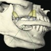
16. Sinus architecturePneumatized sinuses in most patients do not permitthe placement of implants posterior to the cuspid – 1stpremolar region. As a result, A-P spread is insufficient to fabricate an implant supported prosthesis.
17. Fixed vs Removable Masticatory Functionv No difference in chewing efficiency
18. Fixed vs Removable Patient Compliancev Fixed prostheses are very difficult to clean properlyv If the patient has difficulty manipulating hygiene aids they are best suited for a removable overdenture
19. Cost: Fixed vs Removable Overdentures Fixed is double to triple the cost of implant assisted removable overlay dentures.Most expensive Least expensiveFixed prosthesis Implant supported Implant assisteda) More implants overlay dentures overlay denturesb) Time consuming a)More implants a)Fewer implantsand costly to b)Bars need to be milled b)Less labor formanufacture which adds to the manufacture cost of manufacture.
20. Implant assisted overlay dentures Implant assisted with resilient attachments posteriorly Advantages a) Phonetics b) Hygiene access c) Favorable biomechanics d) Better lip contours e) Cost
21. Prosthodontic Designs and Procedures
22. Four Implant Assisted Overlay Denture l Preferred choice in most patients l Implant assisted designs must be used l If A-P spread is adequate and length of implants is adequate, a palateless denture is permitted
23. Implant assisted design developed at UCLA The anterior two implants should be 12-20 mm apart ! This segment is restored with a Hader Barv ERA attachments are positioned adjacent to the distal implants. This attachmentpermits the overlay prosthesis to be compressed into the mucoperiosteum in theextension areas when posterior occlusal forces are applied to the overlay denture. As aresult, the denture bearing tissues absorb the posterior occlusal forcesv This combination of attachments provides excellent retention while at the same time isbiomechanically sound (Williams et al, 2003).
24. Maxillary Retention MethodsRetention of maxillary implant overdenture bars of different designs.Williams BH, Ochiai KT, Hojo S, Nishimura R, Caputo AA.PURPOSE:The purpose of this study was to evaluate the initial retention characteristics of 5 implantmaxillary overdenture designs under in vitro dislodging forces.MATERIAL AND METHODS:A simulated edentulous maxilla was fabricated with 4 screw-type 3.75 x 13-mm implantsanteriorly. Five overdenture designs with the following attachments were evaluated: 4plastic Hader clips with an EDS bar; 2 plastic anterior Hader clips with an identical EDSbar; 2 Hader clips with 2 posterior ERA attachments; 3 Zaag attachments on a bar; and4 Zaag attachments with no bar. Overdentures were fabricated with full palatal coverage.Each design was subjected to 10 consecutive retention pulls on a universal testingmachine. Data were subjected to analysis of variance and t tests to determinedifferences.
25. RESULTS:The highest average value after 10 pulls was 19.8 lb for the combination ERA and Haderclip design. The lowest retentive values were recorded for the 2 and 4 Hader clipdesigns (5.08 +/- 0.89 lb and 5.06 +/- 0.67 lb, respectively). Retention decreased overthe course of consecutive pulls for all designs, especially for the most retentive designs.The smallest retention decrease occurred with the least retentive designs. 26 24 Retentive Force (lb) 22 20 18 16 14 12 10 8 T h e i T h m e 6 a g e c i m a g a e n 4 c n a T ot n h b n e e o i di t m T 2 s pl a y b e d is a g e c h e i m T h e T h i e 0 ERA/Had 4 Zaag dir 3 Zaag/ bar 4 Clip 2 clip Locator Snap Snap/bar
26. Implant Supported vs. Implant Assisted The distance Factors Determining between the Implant Supported vs. Assisted-AP of the center Spread most anterior 4 Implant Assisted Overlay Denture VS. implant and the 4 Implant Rotation” “Axis of Bar Design-Axis distal of the most ERA of RotationHader Attachment posterior implant Implant Assisted Clip Overlay Denture Relatively Favorable
27. Implant Supported vs. Implant Assisted The distance Factors Determining between the Implant Supported vs. Assisted-AP of the center Spread most anterior 4 Implant Assisted Overlay Denture VS. implant and the 4 Implant Rotation” “Axis of Bar Design-Axis distal of the most ERA of RotationHader Attachment posterior implant Implant Assisted Clip Overlay Denture Relatively Favorable
28. Why are implant assisted designs preferred when only four implants are placed? v To minimize the possibility of implant overload and implant failure v At UCLA when implant supported designs were used the distal implants often failed after 3-6 years of service v When we switched to implant assisted designs the failures of posterior implants ceased. Implant supported design Implant assisted design vs
29. Implant Overload – Implant Supported Designs Overlay Dentures in Edentulous Maxilla Four implanted supported overlay dentures with nonresilient (Hader) attachments (arrows) and distal cantilevers Patients # Implants Followup Failures Position Time of of failed failure implants 10 40 5-12 yrs. 4 all distal 39-73 mths.***Failures were attributed to implant overload, causing lossof bone around the implants and eventual implant failure.
30. Four implant supported design Case Report l Hader bar attachments had been placed adjacent to the distal implantsAfter 4 years of use the retention barfractured . Further exam revealed that thedistal implant had lost lost osseointegration.
31. Four implant assisted designImplant placement v Implants should exit at the ridge crest v The two anterior implants should be a minimum of 20 mm apart (measured from the center of the implants) v The posterior implants should be placed as posteriorly as possible to maximize A-P spread. At least 20 mm Maximize A-P Spread ! Implant position in this patient is ideal.
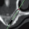
32. Four implant assisted design* Implant angulation! The two anterior implants exit the mucosa at the ridge crest. This is ideal angulation because the bar fabricated will not distort the palatal contours. *Ucla design !The labial inclination of the two anterior implants is of little consequence with respect to the application of occlusal load since this will be an implant assisted design.
33. Four Implant Assisted Overlay Denture*! These two anterior implants are in ideal position.!The distance between the two implants is sufficientfor placement of two clips. As a result the denturewill be more stable and the rate of clip wear will bereduced. *Ucla design
34. Four Implant Assisted Overlay Denture* ! Implant position *Ucla design! These two anterior implants are too close together andwill allow for the placement of only one Hader clip.! Insufficient A-P spread This implant arrangement will result in a less stable denture and the anterior clip will wear more rapidly than if two clips had been possible.
35. Four Implant Assisted Design* Implant angulation ! Note the palatal position and angulation of these implants.!In such a patient it will be difficult to design a barwhich does not distort palatal contours and impair thetongue space. *Ucla design
36. Four Implant Assisted Overlay Denture*! Implant position ! Good A-P spread ! Sufficient space between the two anterior implants
37. Four Implant Assisted Design* Implant angulation and positionPosition and angulation of theimplants in these patients areunfavorable but with a tissue barthese discrepancies can beovercome.
38. Four Implant Assisted Design* Unfavorable implant angulationProblemsv Unfavorable biomechanicsv Increased cost of the laboratory Short narrow diameter implant procedures used as a posterior stop.
39. Four Implant Assisted Design* Implant angulationv The anterior two implants are too close together and angled excessively tothe labial.v The anterior portion of the “Hader” bar is placed palatal to the implants andthe portion over the implants is tapered to allow for proper positioning of thedenture teeth.v Palatal positioning of the bar allows placement of two “Hader” clips.
40. Four Implant Assisted Denture Implant PlacementSurgical templates, generally a duplicate of theexisting denture, will ensure proper implant positionand angulation.
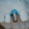
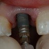
41. Four Implant Assisted Overlay Denture Healing Abutments ! Tapered configuration prevents the mucosa from collapsing over the fixture upon removal of the abutment facilitating impressions, verification of metall Tapered healing frameworks etc. abutments with polished surfaces are favoredl Smooth surfaces retain less plague facilitating more rapid mucosal healing
42. Four Implant Assisted DesignImpression copings ! Transfer type (closed tray ! Pick up type (open tray)
43. Impressionsv Border mold with dental compound in order to develop proper thickness and extension of the labial-buccal flange.v The posterior palatal seal area need not be recordedv Materials used for corrected impression v Polysulfide v Light body polysulfide is preferred by many prosthodontists impressions of the edentulous arch because of its flow and minimal tissue displacement v Permitted when the pickup (open tray) impression copings are linked and are designed to remain in the impression when removing the impression from the mouth v Polysiloxane v Canbe used with transfer type copings because of its rigidity and better accuracy
44. Rubber Base (Polysulfide)v Advantages v Good edge strength v Moderately inexpensive v Optimal flow v Minimal tissue displacementv Disadvantages v Does not recover well from deformation upon removal v The material must be held still during the impression making procedure because the material does not have a snap set v Requires carefully fabricated custom trays that ensure uniform thickness of the impression material v Unpleasant odor v Stains clothing
45. SiliconesAdvantages v More accurate than polysulfide v Less polymerization shrinkage v Low distortion v High tear strength v Fast recovery from deformation v Short working times (3-5 minutes for addition reaction types and 5-7 minutes for condensation types) v Can be used in a stock tray v Available in both hydrophilic and hydrophobic forms v Available in automixing devices v Multiple pours are possible if poured within 1 weekDisadvantages v More expensive compared to polysulfide v Displaces tissues
46. Impressions with linked “pick up” type impression copings and polysulfide!Pickup impression copings are secured tothe analogues imbedded in the preliminarycast with long guide pins.
47. Impressions with linked “pick up” type impression copings and polysulfide!Impression copings are linked togetherwith resin.
48. Impressions with linked “pick up” type impression copings and polysulfideA thin separating disc is used to separate each of thecopings from one another. These will later be lutedtogether on the patient.
49. Impressions with linked “pick up” type impression copingsand polysulfideThe impression copings are blocked out with baseplate wax.The top of the guide pins are left exposed,
50. Impressions with linked “pick up” type impression copings and polysulfide v Separating medium is applied to the cast v The tray is fabricated of tray resin taking care to expose the tops of the guide pins
51. Impressions with linked “pick up” type impression copings and polysulfide v The long guide pins are removed. v The screw access holes are widened to ease insertion of the tray v The portion over the screws is thinned to minimize the bulk of the tray
52. Impressions with “pick up” type impression copings and polysulfidev Border molded impressions made with a custom tray need to be made in order to record the thickness, contour and length of the buccal and labial flange of the overlay denture.v Custom impression tray must be terminate 1-2 mm short of the depth of the vestibule. !The posterior palatal seal area need not be displaced and recorded because a palateless denture is being fabricated.
53. Impressions with “pick up” type impression copings and polysulfidev Once the border molding is completed the impression copings are secured to the implantsv The impression copings are connected together with cyanoacrylate or Duralayv The impression is corrected with a poly sulfide impression material Note: Since this is to be a palateless denture, the posterior palatal seal area was not recorded in the usual fashion.
54. Impressions with “pick up” type impression copings and polysulfidev Once the border molding is completed the impression copings are secured to the implantsv The impression copings are connected together with cyanoacrylate or Duralayv The impression is corrected with a poly sulfide impression material Note: Since this is to be a palateless denture, the posterior palatal seal area was not recorded in the usual fashion.
55. Impressions with “pick up” type impression copings Fixture analogues are attached and the impression is boxed and poured in the usual fashion
56. Impressions with transfer type impression copings and polysiloxane v If transfer type impression copings are used we recommend making the corrected impression with a polysiloxane because of its rigidity and accuracy. v Before use these copings must be carefully inspected for nicks and indentations that may prevent appropriate seating into the completed impression.
57. Impressions with transfer type impression copings and polysiloxanev This impression was made with transfer type copingsv It was border molded with dental compound and corrected with polysiloxane
58. Master castv Note land area. This is useful if a land indexed matrix is used to develop the contours of the bar.v Fixture analogues were imbedded in this cast.v Healing abutments of sizes equivalent to those in the patient were secured in preparation for fabrication of a record base.
59. Record Bases The cast undercuts and those around the healing abutments, are blocked out with wax, separating medium is applied and the record base completed with tray resin.
60. Face bow transfer record is made and the maxillary cast is made
61. Centric relation records! Centric relation records are made following a face bow transfer and mounting of the maxillary cast.
62. Trial dentures! At try in the vertical dimension of occlusion and the centric relation record need to be verified, and a protrusive record made and transferred to the articulator.! The teeth are arranged to achieve bilateral balanced occlusion
63. Land based dental matrix v The land must be 4-5 mm wide if the matrix is to be sufficiently stable. v The matrix is made of silicone putty.
64. Land based dental matrix v Thedenture teeth are attached to the silicone template with sticky wax.
65. Land based dental matrixv Thetemplate positively engages the cast during fabrication of the tissue bar
66. Alternative methodl Occlusal index is made and is mounted on the lower member of the articulator
67. Tissue Bar Fabrication! The path of insertion is determined with a surveyor. Beware of undercuts associated with the alveolar ridge anteriorly.
68. Tissue Bar FabricationUcla abutments ! The UCLA abutment technique is favored because there is a lack of interocclusal space in most patients. ! Fabrication begins by attaching a UCLA abutment to a fixture analogue with a guide pin and coating guide pin and the top of the UCLA abutment with pattern resin. ! Abutment–pattern resin units are attached to fixture analogues imbedded master cast. Non-hexed UCLA abutments are used.
69. Tissue Bar Fabrication ! The Hader bar is used anteriorly, determines the axis of rotation and provides retention. ! The metal housings will eventually become imbedded within the denture base (arrow). ! Note that in cross section the bar is circular, thus permitting free rotation around the axis of rotation.
70. Tissue Bar Fabricationv The Hader bar segment is shaped to the proper contour and length and attached to the anterior implant abutments consistent with the path of insertion and the planned axis of rotation using a specially prepared surveyor rod.v The bar should not touch the tissues of the alveolar ridge
71. Tissue Bar Fabricationv Extracoronalresilient attachments (ERA) are connected to the distal portions of the tissue barv TheERA attachments provide retention and allow the prosthesis to rotate around the “Hader” bar anteriorly when posterior occlusal forces are delivered to the overlay denture
72. Tissue Bar Fabrication! The female portion of the attachment should be secured to the posterior implants with a surveyor.! The path of insertion of the of the Hader bar attachment and the ERA attachments should be identical.! The bar must fit within the confines of the denture base and not distort the palatal contours.
73. Tissue Bar FabricationHader bar is parallel to the occlusal plane and perpendicular tothe midline The ERA attachment should be 1-2 mm above the tissues
74. Tissue Bar Fabricationv Hader segment should be perpendicular to the midline andparallel to the occlusal plane. This will ensure a pure rotationaround the Hader bar when occlusal forces are generatedposteriorly. ! Wax contours are completed making sure there is proper hygiene access ! Space should be sufficient to accept a prophy brush
75. Tissue Bar Fabricationv The anterior portions of the bar is often tapered toaccommodate the anterior denture teeth.v Note the hygiene access beneath the bar andbetween the implants
76. Tissue Bar Fabrication Completed patternv Note the hygiene access between the implants and underneath the barv Note that the ERA attachment is 1-2 mm above the level of the tissuev The tissue bar has been tapered so as to accommodate the proper positioning of the denture teethv At this point the pattern can either be invested and cast or scanned and milled
77. Tissue Bar FabricationFinished casting. Note the hygiene access. If proper hygieneaccess is not provided the gingival tissues will likelyhypertrophy and envelop the tissue bar and the ERAattachments.
78. Tissue Bar FabricationCompleted barv The Hader segment is perpendicular to the midline and parallel to the occlusal plane. It serves as the axis of rotationv The path of insertion for the Hader bar and ERA attachments are identical
79. Tissue Bar Fabrication Completed bar! Note hygiene access beneath bar and between the implants
80. Tissue Bar Fabrication Completed bar ! Metal housing for the ! ERA attachment ERA attachment! Metal housing for Hader clips
81. Tissue Bar Fabrication Completed barThe bar must fit passively, otherwise it must be sectioned and soldered
82. Processingv Prior to processing a metal casting is fabricated toprovide rigidity for the overlay denturev The metal reinforcement framework is secured inits proper position
83. Processingv The metal housings for the attachments are secured to thetissue bar. The black processing attachment is used for theERA .v The tissue bar is blocked out with plaster.
84. Insertion of attachmentsThe processing attachmentis removed with theprovided tool and replacedwith an ERA of light tomedium retentive value.
85. Finished prostheses with attachments insertedIn these cases the housings for the attachments areincorporated within the cast metal of the framework.
86. Delivery Sequence v Attach the tissue bar with gold alloy screwsv Torque the screws to 20 N/cm but no more
87. Delivery Sequence! Secure the tissue bars to the implants! Pressure indicating (PIP) used to eliminate areas of excessive tissue displacement! Check borders with disclosing wax! Clinical remount
88. Delivery SequencePressure indicating paste (PIP) is used to identify and removeareas of tissue displacement during insertion, removal and whenocclusal forces are generated anteriorly. Note the undercut associated with the denture border on the right side (arrow). This must removed to avoid abrading the tissues in this area during insertion and removal of the prosthesis.
89. Delivery SequenceDisclosing wax is used to verify the length andthickness of the denture borders
90. Delivery Sequence Clinical remountPurpose To Correct for the fact that: ! The completed prosthesis seats more accurately than record bases ! Accommodate for errors made during the making of centric relation records
91. Completed Overlay Denture Opposing Fixed Hybrid Prosthesis This maxillary overlay denture is opposed by a fixed hybrid prosthesis. Note the hygiene access beneath the mandibular prosthesis and between the implants.
92. Completed maxillary overlay opposing mandibular overlay• Clips need to changed about every 12-18 months• Denture teeth wear out 7-12 years• Tissue bars wear out 12-15 years.
93. Four implant assisted overlay denturesProblems ! Fracture of acrylic resin overlying the bar ! Wear of denture teeth ! Wear of the tissue bar
94. Problems:! Resin crazing and fracture
95. ProblemsSolution: • Metal reinforcement – Prevents flexure of the prosthesis and crazing and subsequent fracture of acrylic resin overlying the bar
96. ProblemsThe resin is too thin (arrows) overlaying the tissuebar in this case. As a result the resin in this regionwill be susceptible to fracture. The solution is tocover the tissue bar with resin
97. Preventing Resin FracturesMetal frameworks that cover the tissue bar! When lack of inter occlusal space results in resin covering the tissue bar to be excessively thin! Bruxers and clenchers! Brachycephalics! Opposing natural dentition
98. Metal framework designs Metal housings for ERA and Hader attachments have been incorporated within the cast metal framework
99. Bruxers and Brachycephalicsv Patient with brachycephalic facial form with evidence of chronic buxism presented with edentulous maxilla opposing dentate mandiblev Implants were used to restore posterior occlusion in the right mandible
100. Bruxers and BrachycephalicsCompleted prosthesesv Occlusion – Bilateral balance
101. Lack of Interocclusal Space ! Incorporate the metal housings within the metal framework
102. Excessive occlusal wearA B C Wear of denture teeth with implant assisted overlay dentures. Figures A, B,and C (12D years of wear) represent an overlay denture opposing a fixed hybrid prostheses with resin composite denture teeth. Figure D (21 years of wear) represents an overlay denture opposing natural dentition.
103. Excessive occlusal wearAmalgam stops ! These have been relatively ineffective. Note the wear around the amalgam stops.
104. Excessive occlusal wearMetal occlusals ! Chrome cobalt ! Gold occlusals
105. Excessive occlusal wearFifty-five y/o male, presented with a spark-erosion implant supported overlaydenture done delivered in the 80’s. Further tooth loss in the mandibularposterior quadrants were replaced with implant-supported FPD.Exam revealed the maxillary posterior denture teeth to be worn down excessively by theopposing mandibular implant-supported FPD (porcelain occlusals). Solid centricocclusal contacts were found only on anterior teeth. Lack of posterior contact resulted inrecurrent fractures and loss of anterior denture teeth associated with the overlaydenture. The precision of fit between the metal substructure of the overlay denture andthe spark erosion milled bar remained quite good.
106. Excessive occlusal wearA new overlay denture was fabricated using the existing metal substructure.
107. Excessive occlusal wearSolution:Custom gold onlay on the maxillary posterior functional cuspsFunctional cusps were prepare and waxed up toachieve proper contour and occlusal contacts.
108. Excessive occlusal wearv Gold onlay restoring the posterior quadrants were cemented.v Occlusion was refined. Occlusion was bilateral balance
109. Excessive occlusal wear Completed prosthesisLessons Learnedv The probability of rapid and excessive occlusal wear when resin denture teeth are opposed by natural dentition or restorations fabricated with porcelain occlusal surfacesv Custom gold occlusals will reduce the rate of wear and maintain the VOD and solid occlusal contacts.v Osseointegrated implants outlasts the prosthodontic material. Maintenance and replacement costs need to be discussed during initial consult.
110. Excessive occlusal wear Opposing mandibular dentition was restored with porcelain occlusal surfaces. Gold occlusals were used to restore maxillary lingual cusps to minimize wear and prevent loss of VDO.
111. Excessive wear of theHader bar and ERA attachments This implant assisted tissue bar was fabricated of type V gold alloy. It has been in function 21 years. Excessive wear of both the Hader bar and ERA female attachments has made the overlay denture nonretentive.
112. Excessive wear of the Hader bar and ERA attachmentsv Note the excessive wear of the ERA attachment (a) as compared to a newly fabricated tissue bar and attachment (b). This attachment has become nonretentive. This represents 21 years of wear. a b
113. Excessive wear of the Hader bar and ERA attachmentsa bv Note the excessive wear of the Hader bar (a) as compared to a newly fabricated Hader bar (b). This tissue bar has become nonretentive. This represents 21 years of wear.
114. Other implant assisted designs“O” ring type attachments Used when • When implants cannot be splinted together !Non parallel implants results in uneven wear of the attachments and loss of retention.
115. Other implant assisted designs “O” Ring Attachments are used when:! Solitary implants! Implants are so far apart that they cannot be splinted together! Implants are short! Implants in poor quality bone where there is high risk of implantoverload
116. Other Implant Assisted Designs Maxillectomy Defects Note the rests on the bar. The axis of rotation pass through these rests and permits the ERA attachments to function in a way for which they were designed.
117. Other Implant Assisted Designs Patient presents with a repaired bilateral cleft of the lip and palateNote:! The premaxillary segment is missing! The cleft has not been reconstructed with a bone graft! The profound Class III jaw relation !Three implants were placed on each side of the cleft.
118. Bar design uses ERA attachment anteriorly and Hader attachments posteriorlyNote:v The extreme class III jaw relation and the lack of anterior supportv The implant bar does not cross the cleft. These segments move independent of one another during functionv The bar is designed to accommodate the anterior extensionv When occlusal loads are applied anteriorly, the prosthesis rotates around the posteriorly situated Hader attachments. The ERA attachments permits the prosthesis to be impacted into the anterior extension area. This design will limit the force delivered to the anterior implants on each side.
119. Bar design uses ERA attachment anteriorly and Hader attachments posteriorlyNote: Implant the implant assisted design of the tissue bar in the mandible. The bar has been configured to be perpendicular to the midline to enable a more pure rotation when posterior occlusal forces are applied to the denture.
120. Other Implant Assisted Designs Completed prostheses Note: v Implant assisted designs were used in both arches v Full palatal coverage to facilitate support in the maxilla v Occlusion is bilateral balance
121. Other Implant Assisted Designs! Similar problem in a normal patient! Premaxilla resorbed but bone available posteriorly for placement of relatively short implants (8 mm in length)! Opposing natural dentition in the mandible! A bar crossing the arch and splinting both sides together would have created an extensive cantilever predisposing the bar and the anterior implants to a high risk of fracture or implant overload and bone loss.! Occlusion was lingualized with bilateral balance ! Note: Some of the fixed in the mandible was redone to idealize tooth contours and the plane of occlusion
122. v Visitffofr.org for hundreds of additional lectures on Complete Dentures, Implant Dentistry, Removable Partial Dentures, Esthetic Dentistry and Maxillofacial Prosthetics.v The lectures are free.v Our objective is to create the best and most comprehensive online programs of instruction in Prosthodontics


 Cement Retention vs Screw Retention
 Angled Implants
 Prosthodontic Procedures and Complications
 Single Tooth Defects in Posterior Quadrants
