Modern RPD design philosophies recognize that removable partial dentures can exert pathologic stress on abutment teeth that can lead to their failure, particularly in extension based RPD’s. This program discusses the impact of rest position, retainer position and design, and indirect retention on the biomechanics removable partial dentures.
Rpd Biomechanics — Course Transcript
- 1. RPD Biomechanics John Beumer III and Ting Ling Chang DDS Section of Removable Prosthodontics UCLA School of Dentistry This program of instruction is protected by copyright ©. No portion of this program of instruction may be reproduced, recorded or transferred by any means electronic, digital, photographic, mechanical etc., or by any information storage or retrieval system, without prior permission.
- 2. RPD Biomechanics Two types of RPD’s Tooth borne Occlusal forces are transmitted to the teeth used as RPD abutments Extension base Occlusal forces are shared between the abutment teeth and the edentulous denture bearing surfaces. As a result these prosthesis move or rotate during function.
- 3. RPD Classification Systems UCLA – Kratochvil system Based on biomechanics Three types Tooth borne Extension base Unilateral Bilateral Periodontal Stabilization Kennedy – Applegate system Based on edentulous spaces
- 4. Kennedy Classification Based on locations and number of edentulous areas Class I – Bilateral edentulous areas located posterior to the remaining teeth Class II – A unilateral edentulous area located posterior to the natural teeth Class III – A unilateral edentulous area with natural teeth both anterior and posterior to the area Class IV – A single but bilateral (crossing the midline) edentulous area located anterior to the remaining natural teeth Modification spaces Edentulous areas other than those determining the main classes are modification spaces and are designated by the number of spaces present Class IV has no modifications. If more than one space is present in the dental arch it would fall into one of the other classifications
- 5. Forces acting on Removable Partial Dentures Vertical (dislodging) Forces of gravity in maxillary RPD’s Sticky foods Vertical (seating) Forces of occlusion Horizontal (lateral) During the chewing cycle The object of RPD design is to counter these forces without stressing the abutment teeth and the edentulous soft tissue denture bearing surfaces beyond their physiologic tolerance.
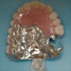
- 6. Types of Removable Partial Dentures Tooth borne Abutment teeth border all edentulous areas Functional forces are transmitted through the abutments to bone It functions like a fixed partial denture Courtesy Dr. G.E. King Courtesy Dr. G.E. King
- 7. Types of Removable Partial Dentures Tooth borne – Essentials of the design Rests on molar teeth should be placed in the center of the tooth Guide planes as parallel as possible Enhance stability (bracing) unilaterally and bilaterally (cross arch stabilization) to resist lateral forces Enhance retention
- 8. Types of Removable Partial Dentures Tooth borne The removable partial denture should provide greatest possible bracing ie. stability (resistance to lateral forces), and support for all the teeth remaining in the arch Ideally and when the occlusion permits molar rests should be extended into the middle of the teeth
- 9. Tooth Borne RPD – Molar rests When the rest is placed on a marginal ridge of a molar, the bone associated with th root on that side becomes overloaded When the rest extends into the center of the tooth the forces are more equitably distributed in the alveolar bone Photoelastic model
- 10. Types of Removable Partial Dentures Tooth borne – Rests When the rests extend to the middle of the tooth the forces are directed down the long axis of the abutment (arrows)
- 11. Types of Removable Partial Dentures Tooth borne- Rests When the rest extends to the middle of the tooth the forces are directed down the long axis of the tooth We confine the rest on premolars to either the mesial or distal side of the tooth or both depending upon the prognosis of the posterior molar. Extending the rest across the transverse ridge may weaken the tooth Courtesy Dr. G.E. King
- 12. Types of Removable Partial Dentures Tooth-Mucosa borne (extension base) Exhibits one or more edentulous areas which are not bordered by abutment teeth Functional forces are shared by both the abutment teeth and denture bearing surfaces in the extension. Unilateral Bilateral
- 13. RPD Biomechanics Extension base RPD’s Anterior extension RPD Posterior extension RPD
- 14. RPD Biomechanics Extension base RPD’s This patient presents with both anterior and a posterior edentulous extension areas Courtesy Dr. A. Davodi
- 15. Extension Based RPD’s Challenge Mucosal bearing surfaces are compressible. Therefore RPD’s are displaced and move during function. Designs must anticipate these movements of the RPD during function to prevent overload and loss of the abutment
- 16. Extension Based RPD’s Amount of movement is dependent upon: The surface area of the mucosal support area The thickness and compressibility of the supporting mucosa The adaptation of the denture base to the tissues of the extension base Refinement of the occlusal factors (distal extension RPD’s) Anterior guidance – Centric only contact posteriorly
- 17. Extension Based RPD’s With improper designs movement of the denture base during mastication or parafunction is destructive to the underlying bone and soft tissue
- 18. Extension Based RPD’s If the denture base is underextended the alveolar ridge will rapidly resorb During mastication or parafunction (clenching and bruxing) the periosteum is compressed, the underlying bone subjected to stress and strain, and a resorptive remodeling response is provoked. An extension base RPD of the Mandible must cover the buccal shelf and the retromolar pad
- 19. Extension Based RPD’s Amount of movement is dependent upon: The surface area of the mucosal support area The compressibility of the bearing surface tissues Therefore, we must maximize the coverage of the edentulous extension area with fully extended impressions. Two methods: Altered cast impressions Fully extended impressions with a custom tray
- 20. The pad contains glandular tissue, loose areolar connective tissue, the lower margin of the pterygomandibular raphe, fibers of the buccinator, and superior constrictor and fibers of the temporal tendon. The bone beneath does not resorb secondary to the pressure associated with denture use. It is one of the two primary support areas of the mandible . Extension Based RPD’s – Retromolar Pad One constant, relatively unchanging structure on the mandibular denture bearing surface is the retromolar pad (dotted line).
- 21. Extension Based RPD’s – Buccal Shelf Boundaries of the buccal shelf: The external oblique line and the crest of the alveolar ridge (area within the dotted lines). The buccal shelf is a prime support area because it is parallel to the occlusal plane . It is composed of dense cortical bone and is relatively resistant to resorption. Masseter groove area Buccinator limits the extension in this area
- 22. Extension Based RPD’s Amount of movement is dependent upon: The surface area of the mucosal support area Therefore, we must maximize the coverage of the edentulous extension area with fully extended impressions Altered cast impressions Fully extended impressions with a custom tray
- 23. Extension Based RPD’s Amount of movement is dependent upon: The surface area of the mucosal support area The thickness and compressibility of the supporting mucosa The adaptation of the denture base to the tissues of the extension base Refinement of the occlusal factors (distal extension RPD’s) Anterior guidance – Centric only contact posteriorly
- 24. Extension Based RPD’s Amount of movement is dependent upon: The surface area of the mucosal support area The thickness and compressibility of the supporting mucosa The adaptation of the denture base to the tissues of the extension base Refinement of the occlusal factors (distal extension RPD’s) Anterior guidance – Centric only contact posteriorly Maximize the surface area and cover key anatomic structures with altered cast impressions
- 25. Extension Based RPD’s Amount of movement is dependent upon: The surface area of the mucosal support area The thickness and compressibility of the supporting mucosa The adaptation of the denture base to the tissues of the extension base Refinement of the occlusal factors (distal extension RPD’s) Anterior guidance – Centric only contact posteriorly This practice will reduce the lateral forces delivered
- 26. Extension Based RPD’s Axis of rotation (fulcrum line) Axis of rotation (fulcrum line) is determined by the position of the rests adjacent to the edentulous extension area. The axis runs through the deepest portion of posterior rests
- 27. Extension Based RPD’s In a posterior tooth the rotation occurs through the depest portion of the rest . Therefore this portion of rest should be contoured as a half sphere We develop this portion of the rest with a #6 or a #8 round burr Proper rest contour
- 28. Extension Based RPD’s When and occlusal force is applied in the denture base extension region: The prosthesis rotates towards the mucosa on the extension base side of the axis of rotation (fulcrum line) All parts of the RPD framework anterior to the axis of rotation (fulcrum line) rotate away from the dentition
- 29. RPD Biomechanics Extension base RPD’s Fulcrum line (axis of rotation) When and occlusal force is applied: The prosthesis rotates towards the mucosa on the extension base side of the fulcrum line All parts of the RPD framework on the dentate side rotate away from the dentition
- 30. RPD Biomechanics Extension base RPD’s Fulcrum line (axis of rotation) (dotted line) When and occlusal force is applied: The prosthesis rotates towards the mucosa on the extension base side of the fulcrum line All parts of the RPD framework on the dentate side rotate away from the dentition
- 31. RPD Biomechanics Extension base RPD’s By changing the position of the rests you idealize the forces delivered to the extension areas and minimize the movement of the extension base Most favorable support in the extension areas is provided when the forces are delivered at right angles to the edentulous bearing surfaces Edentulous extension area
- 32. RPD Biomechanics Extension base RPD’s By moving the rest towards the mesial the forces are applied more vertically in the edentulous extension areas As a result there is less rotation around the axis of rotation (fulcrum line) Edentulous extension area
- 33. RPD Biomechanics Extension base RPD’s Fulcrum line (axis of rotation) Note the rest on the cingulum of the left cuspid. The rest was positioned here because the premolar was periodontally compromised. Note that the proximal plate on the mesial of the premolar provides reciprocation for the retainer as well as stability Advantages: Axis point is lower on the tooth The rest is further away from the edentulous bearing surface resulting in the occlusal forces delivered in a more vertical direction to the extension base
- 34. RPD Biomechanics Extension base RPD’s Fulcrum line (axis of rotation) The RPD rotates around that portion of the rest that is closest to the extension base area Extension base
- 35. RPD Biomechanics Extension base RPD’s Fulcrum line (axis of rotation) Courtesy Dr. A. Davodi
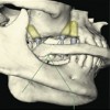
- 36. RPD Biomechanics Extension base RPD’s Fulcrum line (axis of rotation) for an anterior base extension RPD
- 37. RPD Biomechanics Extension base RPD’s This patient presents with both anterior and a posterior edentulous extension areas Therefore, depending on the anterior arch form, there will be two axis of rotation as shown depending whether the patient is incising with the anterior teeth or chewing with the posterior teeth Courtesy Dr. A. Davodi
- 38. RPD Biomechanics Extension base RPD’s Fulcrum line (axis of rotation) The tip of the rest on molar is contoured in a half circle This permits a proper rotation around the axis of rotation
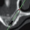
- 39. RPD Biomechanics Extension base RPD’s Position of the retainer From occlusal view, the retainer is placed at the point of greatest mesial-distal curvature of the tooth If the retainer is placed behind the greatest curvature the retainer will move forward during function and torque the tooth and loosen the retention Extension base Point of greatest mesial distal curvature
- 40. Extension Based RPD’s Indirect retention When a dislodging force is applied the prosthesis will rotate around the posterior retainers. The cingulum rest stabilizes the framework during swallowing and tongue thrusting
- 41. Extension Based RPD’s Retainers placed anterior to the axis of rotation in an extension situation should not be in undercuts since that portion of the RPD framework will lift the abutment when a vertical load is applied posteriorly in the extension area. Such retainers should be placed at the height of contour
- 42. Extension Based RPD’s This retainer serves two purposes Retention – Frictional In the event the distal molar is lost This retainer can be slightly can be slightly recontoured and bent to engage the undercut of the canine
- 43. Forces acting on Removable Partial Dentures Vertical (dislodging) Forces of gravity in maxillary RPD’s Horizontal (lateral) During bruxing Vertical (seating) Forces of occlusion (clenching) The object of prosthesis design is to counter these forces without stressing the abutment teeth and the supporting soft tissue denture bearing surfaces beyond their physiologic tolerance.
- 44. Requirements of a Removable Partial Dentures Design based on : Support Rests Major connectors Denture bases Stability (bracing) Minor connectors Proximal plates Rigid portions of retainers Lingual plates Denture bases Rests Retention Direct retainers
- 45. Requirements of a Removable Partial Dentures Provided by : Support Cingulum rests Extension base Major connector Retention Direct “ I” bars on cuspids Proximal plates (when parallel) Indirect Cingulum rests on incisors Stability Minor connectors-proximal plates Lingual plate on anterior teeth Cingulum rests Reciprocation Minor connectors Cingulum rests Proximal plates
- 46. Requirements of a Removable Partial Dentures Provided by: Retention Direct “ I” bars Proximal plates Indirect Cingulum rests Stability Minor connectors and proximal plates Lingual plate on molar Rests Support Rests Extension base Major connector Reciprocation Minor connectors Lingual plate Proximal plates
- 47. Requirements of a Removable Partial Dentures Provided by : Retention Direct “ I” bars premolar Circumferential clasp on molar Proximal plates (when parallel) Indirect Cingulum rests on cuspids Mesial rest on premolar Stability Minor connectors-proximal plates Lingual plate on posterior teeth Cingulum rests Support Occlusal rests Extension base Major connector Reciprocation Minor connectors Cingulum rests Proximal plates Lingual plate
- 48. Requirements of a Removable Partial Dentures Provided by: Retention Direct “ I” bars Proximal plates Indirect Rests – incisal and occlusal Lingual plate Stability (Bracing) Minor connectors and proximal plates Lingual plate Incisal rest Buccal “I” bar on molar Support Rests Extension base Reciprocation Minor connectors Lingual plate Proximal plates Buccal “I” bar on molar
- 49. Principles of RPD design Extension base RPD designs must anticipate and accommodate the movements of the prosthesis during function, without exerting pathologic stresses on the abutment teeth Major connectors must be rigid. Occlusal rest must direct occlusal forces along the long axis of the teeth. Guide planes are created to enhance stability and bracing. Retention must be within the limits of physiologic tolerance of the periodontal ligament. Maximum support is gained from the adjacent soft tissue denture bearing surfaces . Designs must consider the needs of cleansibility.
- 50. Preservation of teeth 1. Extension base RPD designs must anticipate and accommodate the movements of the prosthesis during function, without exerting pathologic stresses on the abutment teeth. Clinical significance: If the RPD designs do not conform to this idea there is risk that abutment teeth may be overloaded leading to their premature loss. Rests on the mesial of teeth adjacent posterior extension area Rests on the distal of teeth adjacent to anterior extension area Courtesy Dr. A. Davodi
- 51. Principles of RPD design 2. Major connectors must be rigid Courtesy Dr. A. Davodi
- 52. Principles of RPD design 3. Occlusal rests must be positive and direct occlusal forces along the long axis of the teeth
- 53. Principles of RPD design 4. Parallel guide planes for stability and bracing
- 54. Principles of RPD design 5. Retention must be within the physiologic tolerance of the periodontal ligament RPI system RPA system Wrought wire
- 55. Principles of RPD design 6. Maximum support is gained from the adjacent soft tissue denture bearing surfaces. Altered cast impressions for extension base partial dentures
- 56. Principles of RPD design 7. Designs must consider the needs of cleansibility and food flow patterns.
- 57. Visit ffofr.org for hundreds of additional lectures on Complete Dentures, Implant Dentistry, Removable Partial Dentures, Esthetic Dentistry and Maxillofacial Prosthetics. The lectures are free. Our objective is to create the best and most comprehensive online programs of instruction in Prosthodontics
New Lectures
 Implants and RPDs
John Beumer III DDS, MS
Ting Ling Chang DDS
Robert Faulkner DDS
Ting Ling Chang DDS
Robert Faulkner DDS
 Restoration of Posterior Quadrants and Treatment Planning
John Beumer III DDS, MS
Robert Faulkner, DDS
Kumar C. Shah DDS, MS
Robert Faulkner, DDS
Kumar C. Shah DDS, MS
 Angled Implants
John Beumer III DDS, MS
Allesandro Pozzi, DDS
Allesandro Pozzi, DDS
 Prosthodontic Procedures and Complications
John Beumer III DDS, MS
Robert Faulkner, DDS
Robert Faulkner, DDS