In the past overlay dentures were primarily employed to restore patients with congenital anomalies such as cleft lip and palate and ectodermal dysplasia. These prostheses restored the missing dentition but also were used to restore the vertical dimension of occlusion and provide support for the lip. This program addresses the principles of RPD framework design that are unique to this application. In addition, a discussion of RPD’s using implants for partial support is provided.
Overlay Removable Partial Dentures — Course Transcript
- 1. 13. Overlay Partial Dentures John Beumer III and Ting Ling Chang DDS Division of Advanced Prosthodontics, Biomaterials and Hospital Dentistry UCLA School of DentistryThis program of instruction is protected by copyright ©. No portion ofthis program of instruction may be reproduced, recorded or transferredby any means electronic, digital, photographic, mechanical etc., or byany information storage or retrieval system, without prior permission.
- 2. Overlay Partial Dentures
- 3. Overlay Partial DenturesAdvantages v Maintain tooth support v More favorable biomechanics – Improved crown-root ratio v Maintains bone. In the event of the loss of the root tips bone sites are maintained for implants v Improved esthetics
- 4. Overlay Partial Dentures Overlay RPD’s first used in the 1930’s primarily for the treatment of cleft lip and palate patientsThese patients often presentedwith: v Loss of the premaxilla v Collapse of the posterior segments inhibiting proper eruption of teeth and vertical development of the alveolar ridges v Collapsed vertical dimension of occlusion with excessive interocclusal space
- 5. Overlay Partial Denturesv This is a typical design used in the 1950’s and 60’s. These photos were made in1968. The primary focus was retention.
- 6. Overlay Partial Denturesv By 1991 the patient had lost multiple teeth secondary to periodontal disease and dental caries
- 7. Overlay Dentures and RPD’sLessons learned from this era v Overlay denture abutments are susceptible to caries and periodontal disease v Prevention: v Do not cover the abutments with the acrylic resin of the denture base. Keep acrylic resin away from the tooth tissue junction v Cover the abutments with gold copings v Cover the abutments with the metal framework of the overlay RPD with metal extending beyond the tooth tissue junction v Emphasize oral hygiene v Place a drop(s) of fluoride gel onto the portion of the RPD overlaying the abutment(s)
- 8. Overlay Partial Denturesv Shown is an overlay designedin the 1990’sv The vertical dimension ofocclusion was restored withselective crown placement andan overlay denture.v A gold coping with anattachment had previously beenplaced on the left premolar
- 9. Overlay Partial DenturesCopings on premolars to be These copings will be enclosed within theover-laid to protect these teeth overdenture.from caries. Crown and coping contours were developed based on the survey line made ideal for the partial overdenture.
- 10. Overlay Partial DenturesNote the thickness of the The processed overdenture with the borderlabial flange required tosupport the upper lip molded soft palate obturator. v The gold copings were Attachment covered with metal but note that the coping with the attachment is covered with and the female portion of the attachment is embedded within acrylic resin. v This will predispose these gingival tissues to irritation andThese copings areincorporated within the hypertrophy. Today it isoverlay denture. possible to incorporate the housing for the attachment in the metal framework
- 11. Overlay Dentures and RPD’s Prevention of soft tissue irritation and hypertrophy: v Do not cover the abutments with acrylic resin of the denture base. Keep acrylic resin away from the tooth tissue junctionThese copings are covered with acrylic resin. This willpredispose the gingival tissues to irritation and hypertrophy.Today it is possible to incorporate the housing for the attachmentin the metal framework
- 12. Overlay removable partial denture. Metal housings for ERA and Hader attachments have been incorporated within the cast metal framework of an overlay prosthesis
- 13. Overlay Partial Dentures v Anterior open bite v Fistulas v Soft palate defect
- 14. Overlay Partial Denturesv Note the housings for the Hader clips have been incorporated within the metal framework.v The copings and the tooth tissue junctions have been covered with metalv Such practice, combined with good oral hygiene will ensure that the health of gingival tissues surrounding the copings will be maintained
- 15. Overlay removable partial denture.Occlusal plane discrepancies in the mandible were addressedwith restorations and recontouring enamel surfaces
- 16. Overlay Dentures and RPD’s Designv The lingual portion of the teeth and the tooth tissue junction are covered with metalv Retainers engage undercuts on the buccal surfaces.v All buccal surfaces are blocked out to prevent contact with the acrylic resin
- 17. Overlay RPD’sv Splinted, surveyed crowns with positive rests.v Cover overdenture abutments and tooth tissue junction with metal.v Overdenture abutment coping rounded to permit rotationv Topical fluoride used daily
- 18. Overlay RPD’sCaries preventionv Cover overdenture abutments and tooth tissue junction with metal for improved tissue response.v Topical fluoride use daily for caries controlv Casein based calcium phosphates for remineralization
- 19. Overlay Partial DenturesPremaxilla of this patient hasbeen removed. Note fistulaanteriorly. This must becarefully occluded with gauzebefore making impressions.The maxillary molars wererestored with full veneercrowns to restore the VDO to aproper level. Premolars covered with copings and splinted to the first molars. The prosthesis will overlay these copings. Note the ERA attachments (arrows). These attachments retain the prosthesis but disengage when incisal forces are delivered. The axis of rotation is determined by the distal rests on the first molars bilaterally (arrows).
- 20. Overlay Partial DenturesThis cleft was not bone grafted and therefore theindividual segments move independent of one anotherwhen exposed to the forces of mastication. Thereforecleft segments have not been united prosthodontically.
- 21. Overlay Partial DenturesThe prosthesis inserted. Circumferential claspshave been used. Note the anterior overlay.
- 22. Overlay Partial DenturesThe anterior overlay restores the anterior dentition,provides lip support and obturates the anteriorfistula.
- 23. Implants and RPD’sApplications and rationalev Implants with questionable anchoragev Implants with unfavorable biomechanicsv Replacement for a key abutmentv Supplement existing dentitionv Unanticipated implant failures
- 24. 23 y/o female, S/P auto vehicle accidentv Occlusal plane discrepanciesv Implants in grafted bone • Bone implant interfacev Biomechanics unfavorable • Unfavorable crown root ratios • Unfavorable implant angulation • Linear configuration • Buccal cantilever • Posterior occlusion
- 25. Treatment planning and RPD design concept 1. Splint implants together with a tissue bar 2. Hader bar is designed to consistently follow the axis of rotation of this unilateral distal extension RPD 3. ERA attachment allows 0.4 mm vertical movement before active engagement 4. Implants are designed for retention (from Hader bar and ERA) and stability. Support is also provided by teeth and mucosa 5. Resolve occlusal plane discrepancy
- 26. Tissue bar design Note hygiene access
- 27. Completed overlay RPDFinal finished implant tissue bar Black-colored ERA processing pattern replaced with Orange-colored ERA Yellow Hader clip in the metal housing
- 28. Completed overlay RPD Implant-assisted overlay partial denture provides favorable biomechanics and also offers optimal esthetics for lip/cheek support and replace hard and soft tissue
- 29. Why Implants and RPD in this patient? v Implants were in grafted bone v Implant/restoration ratio unfavorable v Facilitate support, stability, retention v Implants, teeth, mucosa v Esthetic considerations
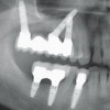
- 30. Unanticipated implant failures52 y/o male, maxillary partially dentate, 6 implants placed
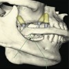
- 31. Unanticipated implant failures4 out of 6 maxillary implants failed to osseointegrate Note buccal inclination
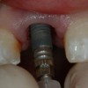
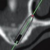
- 32. Unanticipated implant failuresv Two custom abutments with ERA Attachments were made to correct the labial angulation consistent with the path of insertion of the two ERAs of #8 & 9 and the guide planes of the molars Note that implants are used for retention and stability, not support.
- 33. Implant Prosthesis Design Conceptv Implants’ labially inclined angulation were corrected by custom abutment with ERA plastic patternsv Due to loss of the other 4 implants, maximize the support by positive rests on #2, 8, 9, 14v Additional support obtained fro from the palatal connectorv Implants are used for v #8 and 9 are splinted and ERA’s retention (from ERAs). Support and stability are designed to idealize esthetics provided by teeth and mucosa
- 34. Completed overlay RPDv White-colored (the least amount of retention) ERA were used on the implants.v Support provided by the positive occlusal and cingulum rests and the full palatal coverage
- 35. Completed overlay RPD
- 36. Issues in this casev Despite the best effort implants sometimes fail to osseointegratev Prosthodontic contingency plans should be discussed during the initial consult.v Few implants remained with poor angulationv Support, stability, and retention v Positive rests, full palatal coverage
- 37. Unanticipated implant and abutment failuresv Both natural tooth abutments were treated endodonticallyv The right 2nd molar was lost and the 2nd premolar fractured and was extracted
- 38. Unanticipated implant and abutment failures v Eventually the implant and the 1st premolar in left mandible failed. v An implant was placed into the 1st premolar site v An ERA attachment was used and the overlay extended to the left side
- 39. Supplement existing dentitionThis patient is S/P maxillectomy. The right soft palate hasalso been resected. The anterior teeth were lostsecondary to periodontal bone loss. Implants were placedin the residual premaxillary segment.
- 40. Supplement existing dentitionv Implants used to facilitate retention stability and supportv Rests (arrows) are milled into the bar between the implants.v Occlusal forces are directed through the restsv ERA attachments used for retention
- 41. Supplement existing dentition Rests ERAAttachments Note the metal substructure incorporated within the finished prosthesis. It contains the ERA attachments and the occlusal rests.
- 42. Supplement existing dentitionv Note the contours of the obturator. The extension into the velopharyngeal area is about 10 mm in height.v Note the prominent gingival roll posteriorly on the defect side to deflect the cheek away from the biting surfaces Retention: The prosthesis in position. ” Implantselevation of the right Note the ” Lateral wall extension so lateral incisor and cuspid ” Posterior molars lip as to follow the line.
- 43. Replacement for a key abutment
- 44. Replacement for a key abutment
- 45. Overlay Partial Dentures l Anterior open bite l Fistulas l Soft palate defect
- 46. Overlay Partial Denturesv Remaining teeth covered with gold copings.v Implants placed in posterior quadrant.
- 47. Overlay removable partial denture.
- 48. Overlay removable partial denture.Adult with ectodermal dysplasia. This patientpresents with a full spectrum of the side effects ofthis disease complex. She presents with multiplemissing and malformed teeth and an excessiveamount of interocclusal space.
- 49. Overlay removable partial denture. Transitional Removable Prosthesis Phase
- 50. Overlay removable partial denture.Two implants were placed in the maxilla and four inthe mandible. An overlay was planned for themaxilla to restore the VDO, and fixed was plannedto restore the mandibular dentition.
- 51. Overlay removable partial denture.The fixed partial denturefor the mandible wasporcelain fused to metalfabricated with the UCLAabutment.
- 52. Overlay removable partial denture.The fixed prosthesis for the mandible and thetissue bar for the maxilla has been inserted.
- 53. Overlay removable partial denture.The maxillary overdenture. This prosthesis restores the verticaldimension of occlusion and replaces the missing dentition.
- 54. Overlay removable partial denture.The overlay partial denture inserted. Note thedramatic change in esthetics.
- 55. Overlay removable partial denture.Implants can provide cleft patients significant benefit particularly thoserestored with an overlay prosthesis. This patient has retained only themaxillary 2nd molars. The removable partial denture she was wearingrestored the missing dentition and provided support for the lip but lackedanterior support. Implants provided the needed support.
- 56. Overlay removable partial denture.A lateral cephalogram indicated sufficient bone availableanteriorly and implants were place on both sides of the cleft.Note that the premaxilla had been removed previously.
- 57. Overlay removable partial denture. Implants can provideA “Hader bar” and an “O” ring type attachment wereuse to retain the overlay prosthesis. The occlusal surfaces of the mandibular teeth were altered to alleviate occlusal plane descrepancies.
- 58. Overlay removable partial denture.The finished prosthesis in position. The posteriormandibular dentition was restored with a removable partialdenture. The cleft segments had not been grafted togetherand so they were not splinted with the implant apparatus.
- 59. v Visitffofr.org for hundreds of additional lectures on Complete Dentures, Implant Dentistry, Removable Partial Dentures, Esthetic Dentistry and Maxillofacial Prosthetics.v The lectures are free.v Our objective is to create the best and most comprehensive online programs of instruction in Prosthodontics
New Lectures
 Restoration of Posterior Quadrants and Treatment Planning
John Beumer III DDS, MS
Robert Faulkner, DDS
Kumar C. Shah DDS, MS
Robert Faulkner, DDS
Kumar C. Shah DDS, MS
 Angled Implants
John Beumer III DDS, MS
Allesandro Pozzi, DDS
Allesandro Pozzi, DDS
 Single Tooth Defects in Posterior Quadrants
John Beumer III DDS, MS
Robert Faulkner, DDS
Robert Faulkner, DDS
 Prosthodontic Procedures and Complications
John Beumer III DDS, MS
Robert Faulkner, DDS
Robert Faulkner, DDS