Most of palatal defects are caused by the surgical resection of maxillary and paranasal sinus tumors. However, trauma, fungal infections, prolonged cocaine use, and recently osteonecrosis secondary to use of bisphosphonates can also cause substantial boney and soft tissue defects in the palate. This program describes some of the most common tumors, and the methods used for their resection. Defects secondary to trauma, aspergillosis, mucormycosis and osteonecrosis secondary to bisphosphonates are also show and discussed if regards to challenges of faced when attempting to obturate these defects with a prosthesis.
Maxillofacial Prosthetics – Etiology of Palatal and Paranasal Sinus Defects — Course Transcript
- 1. Etiology of Palatal Defects John Beumer III, DDS,MS Distinguished professor emeritus UCLA School of Dentistry All rights reserved. This program of instruction is protected by copyright ©. No part of this program of instruction may be reproduced, or transmitted by any means, electronic, digital , photographic, mechanical etc., or by any information storage or retrieval system, without prior written permission from the authors .
- 2. Table of Contents Tumors of the region Epidermoid Salivary gland Mesenchymal Traumatic defects Fungal infections Osteonecrosis secondary to bisphosphonates Prolonged cocaine use
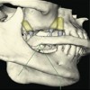
- 3. Tumors of the Region: Resection and the resultant defects Maxillofacial Prosthodontists need to be familiar with the types of surgical resections used to remove various types of tumors found in the upper jaw and paranasal sinuses. Why? Advantages: He/she can better interact with the surgeon The prosthodontist needs to become familiar with the methods of treatment so that he/she can better prepare the patient to deal with the limits of oral function and morbidities imposed by the surgery and adjunctive therapies such as radiation therapy or chemoRT.
- 4. Tumors of the Region: Epidermoid Sinus Palatal Salivary gland Benign Malignant Mesenchymal Benign Malignant
- 5. Methods of Resection Palatectomy Performed transorally. Mucosal incisions are outlined to provide 5-10 mm of normal tissue around the tumor. Bony cuts are made with a power saw or an osteotome. The bony edges are covered with residual mucosa and periosteum if available. Split thickness skin grafts are used to line selected raw tissue surfaces of the defect. Radical maxillectomy Skin incisions (Weber-Fergusson) are used to expose the maxilla to be resected. Oral mucosal incisions are made through the palate and the buccal vestibule. Bony cuts are made through the palate, alveolar ridge, lateral nasal bones, floor of the orbit, malar eminence, pterygoid plates and zygomatic arch. After the remaining soft tissues are detached and removed with the specimen, the raw tissues of the defect are lined with a split thickness skin graft. The skin incision is then closed.
- 6. Methods of resection Radical Maxillectomy A typical resection and defect is shown. Note that the defect is lined with skin. This type of defect is a favorable one because the defect can be used to facilitate the retention, stability and support of the partial denture with obturator . In this patient an orbital an exenteration was also performed .
- 7. Palatectomy These defects are generally quite small compared to radical maxillectomy defects and usually the paranasal sinus partitions are still intact. Dentate patients with such defects are relatively easy to restore if suitable numbers of teeth are present and the defect does not extend into the movable portion of the soft palate. Edentulous patients with such defects are more difficult to restore because the defect cannot be engaged as aggressively compared to a skin lined radical maxillectomy defect, resulting in compromised retention. Such patients stand to benefit from the placement of osseointegrated implants.
- 8. Epidermoid carcinomas Tumors arising from the paranasal sinuses Tumors arising from the palatal epithelium
- 9. Epidermoid carcinomas arising from the paranasal sinuses Initial signs and symptoms : Nasal congestion Nasal infection and bleeding Loosening of teeth and other oral signs Proptosis and swelling around the eye and cheek
- 10. Epidermoid carcinomas arising from the paranasal sinuses Roentgenographic analysis reveal soft tissue masses filling the sinus with advanced lesions demonstrating erosion of the bony walls of the antrum (arrows).
- 11. Epidermoid carcinomas arising from the paranasal sinuses In both these patients the first signs that brought the tumor to the attention of the patient was palatal swelling and loosening of teeth.
- 12. In these three patients, swelling of the cheek and eye region first brought the attention of the patient to the tumor.
- 13. Epidermoid carcinoma arising from the paranasal sinuses These tumors generally require radical maxillectomy sometimes accompanied by an orbital exenteration for tumor removal. Many patients also receive postoperative radiation. Metastasis to the neck is rare and so prophylactic neck dissections are generally not performed.
- 14. Epidermoid carcinomas arising from the paranasal sinuses Defects secondary to radical maxillectomy can be rather large and disfiguring and fabrication of the prosthetic restoration can be quite challenging particularly in the edentulous patient. Osseointegrated implants are strongly recommended in these situations.
- 15. Epidermoid carcinomas arising from the palatal mucosa These tumors tend to stay localized and generally can be removed with partial palatectomy transorally. The defects created are smaller than those created with radical maxillectomy and less disfiguring. Prosthodontic restoration of these types of defects is relatively simple particularly when key teeth are still present. If teeth are not present implants can be placed to retain and stabilize the prosthesis.
- 16. Salivary gland tumors Benign Pleomorphic adenoma Most arise from the minor salivary glands at the junction of the hard and soft palate. They stay localized but require resection with a healthy margin of normal tissue around the tumor. A palatectomy performed transorally is usually sufficient for removal.
- 17. Pleomorphic adenoma Even large pleomorphic adenomas such as this can be removed transorally with a partial palatectomy. Prosthetic obturation is relatively straight forward as long as healthy teeth or bone sites for implants remain.
- 18. Pleomorphic adenoma The surgical defects created are usually confined to the junction of the hard and soft palate. Prosthetic obturation is easily accomplished. Occasionally when the defect extends onto the middle third of the soft palate, leakage of fluids during swallowing may occur sporadically.
- 19. Salivary gland tumors Malignant Adenoid cystic carcinoma These are slow growing but locally aggressive malignant tumors that tend to spread along peripheral nerves and perivascular sheaths. They require very aggressive local resections.
- 20. Adenoid cystic carcinoma Aggressive resections are required for cure. Note how much of the hard palate has been removed in this resection. This combination of hard palate – soft palate defect however is easily obturated prosthetically providing a levator veli palatini remnant is present on the side opposite the tumor resection and teeth or implants are available to retain the prosthesis.
- 21. Adenoid cystic carcinoma Note the recurrence (arrow). This is a common occurrence and patients with adenoidcystic carcinoma often require additional resections. Fortunately however, it is a slow growing neoplasm that remains localized in most patients.
- 22. Mucoepidermoid carcinomas High grade Intermediate grade Low grade Resection of high grade tumors is aggressive often requiring a Weber-Fergusson incision while most low grade tumors can be removed transorally .
- 23. Mesenchymal Tumors Lymphosarcomas Chondrosarcomas Osteosarcomas This patient presented with an osteosarcoma of the hard palate. Note that the tumor has invaded into the floor of the nose and the maxillary sinus (arrow). A transoral partial palatectomy was performed and the defect obturated prosthetically .
- 24. Mesenchymal Tumors This patient presented with an extensive osteosarcoma. A radical maxillectomy and an orbital exenteration was required for tumor removal. The prognosis for such an advanced neoplasm is very poor.
- 25. Other phenomenon causing maxillary defects Trauma Aspergillosis Mucormycosis Osteonecrosis secondary to use of bisphosphonates
- 26. Traumatic Defects A traumatic defect secondary to a gunshot wound Note the poor quality mucosa lining the defect, its irregular shape and the scarring of tissues adjacent to the defect. Only the maxillary second molar remains.
- 27. Traumatic Defects A traumatic defect secondary to self inflicted gunshot wound Note the poor quality mucosa lining the defect, its irregular shape and the scarring of tissues adjacent to the defect. The residual maxillary segments are displaced and the mandibular fragments are misaligned.
- 28. Traumatic Defects These defects are more difficult to restore prosthodontically. Why ? Poor quality mucosa lines the defect Defects are irregular in size and shape Scarring of tissues adjacent to the defect Residual maxillary segments may be displaced Misalignment of the mandibular dentition complicate the occlusal relationships
- 29. Other phenomenon causing maxillary defects Mucormycosis and aspergillosis These are fungal infections that occur in chronically immuno-suppressed patients and those with uncontrolled diabetes. The organisms cause ascending venous thrombosis resulting in necrosis of the involved structures.
- 30. Mucormycosis and aspergillosis Treatment requires extensive resection combined with systemic antifungal therapy. The defects cannot be successfully skin grafted and may be difficult to restore because of scar contraction and the poor quality of the epithelium lining the defect. Immediately postoperative 4 months postoperative
- 31. Mucormycosis and aspergillosis This patient required removal of the orbital contents to control the infection resulting in facial disfigurement.
- 32. Cocaine use This defect was the result of prolonged use of cocaine.
- 33. BRONJ Most of the defects associated with bisphosphonate use are localized as seen here.
- 34. Visit ffofr.org for hundreds of additional lectures on Complete Dentures, Implant Dentistry, Removable Partial Dentures, Esthetic Dentistry and Maxillofacial Prosthetics. The lectures are free. Our objective is to create the best and most comprehensive online programs of instruction in Prosthodontics
New Lectures
 Angled Implants
John Beumer III DDS, MS
Allesandro Pozzi, DDS
Allesandro Pozzi, DDS
 Cement Retention vs Screw Retention
John Beumer III DDS, MS
Robert Faulkner DDS, MS
Robert Faulkner DDS, MS
 Prosthodontic Procedures and Complications
John Beumer III DDS, MS
Robert Faulkner, DDS
Robert Faulkner, DDS
 Restoration of Posterior Quadrants and Treatment Planning
John Beumer III DDS, MS
Robert Faulkner, DDS
Kumar C. Shah DDS, MS
Robert Faulkner, DDS
Kumar C. Shah DDS, MS