This elegant presentation describes the clinical impact of cervical contour, marginal adaptation, and surface smoothness of complete coverage crowns on the health of the gingiva around natural dentition and the peri-implant mucosa circumscribing implant crowns. The clinical impact of over-contouring the cervical portion is dramatically illustrated. The importance and clinical impact of the so-called “S” contour of the cervical region of the crown is shown in detail with numerous clinical demonstrations. This presentation also demonstrates the value of flat, under-contoured cervical surfaces in order to preserve gingival form and allow the gingiva to return to its normal position. The importance of fabricating properly contoured provisional crowns with smooth surfaces, in promoting gingival recovery and health is emphasized. Finally the application of these principles to the design and development of appropriate contours of implant crowns is discussed and described.
Transcript
- 1. Charles J. Goodacre, DDS, MSD Professor of Restorative Dentistry Loma Linda University School of Dentistry This program of instruction is protected by copyright ©. No portion of this program of instruction may be reproduced, recorded or transferred by any means electronic, digital, photographic, mechanical etc., or by any information storage or retrieval system, without prior permission. The Impact of Cervical Contour, Marginal Fit, and Surface Smoothness on the Gingiva & Peri-Implant Mucosa
- 2. The Impact of Cervical Contour, Marginal Fit, and Surface Smoothness on the Gingiva & Peri-Implant Mucosa Charles J. Goodacre, DDS, MSD Professor of Restorative Dentistry Loma Linda University School of Dentistry
- 3. What I Sometimes See Around Ceramic Crowns: Gingiva that is red, edematous, has abnormal form and position, or a combination of the above
- 4. The Effect of Cervical Overcontouring
- 5. The Combined Effect of Cervical Overcontouring & Poor Oral Hygiene
- 6. Consequences Of Cervical Contour During Adolescence
- 7. The End Interference With Normal Cervical Relocation
- 8. Causes of Poor Gingival Responses • Lack of optimal periodontal health before treatment • Less than optimal oral hygiene before, during, and after treatment • Excess trauma during treatment • Contour, fit, and surface smoothness of provisional crowns and definitive crowns
- 9. The Benefit of Optimal Periodontal Health Before Prosthodontic Treatment • Less likely to have adverse gingival response during or after treatment • Better esthetic result because “gingival esthetics” and “tooth esthetics” are both required for an optimal esthetic result (they complement each other)
- 10. Lessons I Learned From Poor Gingival Responses Around Ceramic Crowns
- 11. My Lessons Began In The Early 1970’s
- 12. Example of Overcontoured Crown & Severe Marginal Gingivitis The Lessons Continued For Decades
- 13. Subcrestal Tooth Fracture & Heroic Restoration That Was Not Expected To Last Patient History
- 14. 2-15-1990
- 15. 10-26-2005 2-15-1990
- 16. Lessons Learned From 40 Years Of Developing Crown Contours On Natural Teeth – Benefits of The S-Shaped Cervical Contour Patient Histories
- 17. Preserving papillae between both teeth and implants is difficult when the distance from the interproximal bone to the proximal contact is substantial.
- 18. Good marginal fit, surface smoothness, and the subtleties of the S-shaped cervical contour promote PAPILLA PRESERVATION
- 19. 17-year PO 2014 Crown placement 1997
- 20. Poor Gingival Response To Existing Metal Ceramic Crowns On The Maxillary Central Incisors
- 21. • Oral Hygiene • Fit • Contour • Surface Roughness • Retained Cement • Rough Areas On Teeth Below Crowns • Calculus • Type of Alloy What are the possible causes?
- 22. Periodontal Treatment Improved The Gingiva But Did Not Solve The Problem
- 23. Crowns Removed; Extensive Bleeding; Teeth Reprepared; Provisional Crowns Made With Flat Cervical Contours PermaSeal Resin Glaze from Ultradent applied, light polymerized, and air inhibited layer removed
- 24. Gingival Appearance When Patient Returned For Crown Cementation In Three Weeks
- 25. Prosthodontic Laboratory Procedures Completed by Mr. Satoshi Sakamoto, LLUSD Dental Laboratory
- 26. At Cementation 4 Weeks Postcementation
- 27. December 2013 – 4 year PO
- 28. 4 Weeks Postcementation Pretreatment Why Was There Improvement?
- 29. Why Was There Improvement? • Periodontal surgery (unhealthy tissue removal; calculus / cement removal)
- 30. Why Was There Improvement? • Reduction depth & smooth finish line • Fit, smoothness, & cervical contour of crowns
- 31. Why Was There Improvement? • Slight S-shaped cervical contour
- 32. Multiple Overcontoured Crowns & Marginal Gingivitis The Importance of Cervical Contour Patient History
- 33. Pretreatment Provisional crowns after impression At crown cementation appointment
- 34. Pretreatment Cementation – December 2011
- 35. 9 month PO August 2012 17 month PO May 2013
- 36. 17 months Post-Treatment Pretreatment
- 37. 2 years following placement of the crowns, the post in tooth #9 came loose along with the crown
- 38. There was little coronal tooth structure remaining after the post failure. The treatment options were to re-restore the tooth or extract it and place a dental implant Excess bone-to- proximal contact distance The bone-to-proximal contact distance indicates it would be unlikely to retain papillae
- 39. The root was re-prepared and slightly lengthened. An impression was made for a cast gold post and core A prefabricated post was adapted to the post space and the inside of the crown to serve as a provisional crown Flowable composite resin placed around proximal contacts to help retention
- 40. A cast post and core was fabricated along with a provisional crown
- 41. The post and core was cemented using RelyX Unicem resin cement The mucosa after refining the tooth preparation, making an impression, and cementing the provisional crown The tooth preparation was refined and impression made
- 42. Mucosa around provisional crown at time of new crown cementation Provisional crown removed & cleaned
- 43. New crown
- 44. New crown
- 45. Prior to P&C failure New crown cemented
- 46. New crown cemented 4-month PO
- 47. Using The Contour Principles To Maintain The Gingiva Around Crowns Patient Histories
- 48. 2-Year PO
- 49. Provisional Crown Characteristics That Promote Optimal Gingival Health • Good fit • Flat, undercontoured subgingival surfaces • Smooth, glazed external surface
- 50. Examples of Flat, Undercontoured Cervical Surfaces on Provisional Crowns Their Benefit in Preserving Gingival Form and Position or Allowing Gingiva to Return to its Normal Position
- 51. After use of gingival displacement cord, making an impression, and cementing the provisional crown, the gingival was displaced apically
- 52. The flat, undercontoured portion of the definitive crown ends at the desired location of the gingival margin and normal contours begin at this location.
- 53. Provisional crown after preparation & impression Provisional crown at cementation appointment All-ceramic crown
- 54. Two weeks postcementation
- 55. Provisional Crown & Connective Tissue Graft
- 56. Another provisional crown made after connective tissue graft healing to promote incisal migration of the gingiva.
- 57. Definitive Metal Ceramic Crown
- 58. Three-Year PO
- 59. Provisional crowns with flat cervical contours
- 60. All-ceramic crowns with flat subgingival contours
- 61. 5 years after placement patient was drinking from a bottle and someone bumped her and the bottle chipped crown #9 Hydrofluoric acid etch of porcelain and resin repair
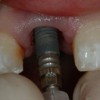
- 62. Cervical Crown Contour & Dental Implants • Applying the principles and procedures used with natural teeth to dental implant treatment
- 63. Subcrestal Tooth Fracture & Heroic Restoration That Was Not Expected To Last “THE REST OF THE STORY” Patient History
- 64. 10-26-2005 2-15-1990
- 65. 8-14-2006 16-year PO
- 66. 2010 20-year PO
- 67. Patient With A Poor Gingival Response To Existing Crowns; Root Resorption On Tooth #10 That Requires Extraction Patient History
- 68. Provisional crown shaped Provisional crown glazed
- 69. Outcome of undercontoured or flat cervical morphology when patient returned for definitive crowns
- 70. 2 weeks Following Crown Placement Gingiva at Crown Placement Appointment
- 71. 4-Years Following Crown Placement
- 72. Patient History Patient who was losing a maxillary central incisor due to root fracture and there was a slightly open cervical embrasure. He wanted an improved esthetic result without gingival discoloration.
- 73. Patient History Congenitally missing tooth #7 with canine in that position. Canine now has root resorption. Extraction & immediate implant placement.
- 74. Restoration of the initial resorption under flap reflection was not successful
- 75. 1-year PO
- 76. Treatments With Less Than Optimal Results And Unfavorable Results That Required Re-Treatment
- 77. Patient History Patient is a smoker and never gets around to oral hygiene. Patient left periodontist after treating the maxillary anterior area due to his inability to meet OH expectations. Caries is a regular event & periodontal disease continues. I am now supervising the loss of teeth with “as much dignity & respect as I can provide”.
- 78. 6-Year PO
- 79. Patient History Congenitally missing maxillary lateral incisors. Distance from interproximal bone to proximal contact was 4 mm on one side and 6 mm on the other. Research has shown that a papilla will only be present about 50% of the time when the distance is 6 mm or more Choquet, 2001
- 80. Note the difference in papilla height between the left and the right and the corresponding bone levels
- 81. 1-Year Postplacement 11-Years Postplacement
- 82. Patient History Traumatic injury resulted in bone & soft tissue loss. Grafting made it possible to place implants but defects still present. Thin biotype & no interdental papillae
- 83. • Interdental papillae can seldom be recreated with thin tissue when the distance from the bone to the marginal gingiva is greater than 4 millimeters Kan, 2003
- 84. Thin Biotype & 6 mm Distance from Bone to Proximal Contact Contour facilitates preservation 7 years PO
- 85. Poor Gingival Response To Existing Crowns Biologic Width Violation: Tooth # 9 Prepared To The Bone Level With Feather Edge Finish Line That Also Produced Overcontoured Crown Patient History
- 86. Sequestrum of Bone Found Mesiofacially
- 87. Provisional Crown At Cementation
- 88. Definitive Crown After Cementation Provisional Crown At Cementation
- 89. After 1 week
- 90. Reason For Improvement • Marginal Fit • Cervical Contour • Surface Smoothness
- 91. One month post-cementation Unfortunately, the favorable response did not last
- 92. Two months PO: Gingiva getting worse
- 93. Tooth #9 will have to be extracted. The fee patient paid for the crown will be applied to the cost of the implant crown.
- 94. After Cementation After 4 Weeks
- 95. Two months PO of first crown One month PO of Implant Crown
- 96. Patient History Implants placed by another practitioner and patient upset with result. I was able to obtain pretreatment photo and radiograph
- 97. Should have considered orthodontic eruption prior to extraction
- 98. Photoshop simulation of incisocervically shorter crowns with gingival colored porcelain
- 99. Provisional crowns made by dental laboratory for clinical trial placement and evaluation
- 100. Trial placement of provisional crowns shows they are too long cervically. The crowns were modified by removing white resin cervically and replacing with pink resin until the correct dimensions and form were developed. They were returned to the laboratory to serve as a guide for fabrication of the definitive crowns
- 101. Definitive Crowns and Maximal Smile
- 102. Patient History Implant placed by a surgeon and then the patient referred to me for restoration. Implant angled facially and no interproximal papillae
- 103. 1.5 year PO 1-month PO
- 104. PATIENT TREATMENT Fracture of root canal treated tooth where there had been some bone loss necessitated extraction of maxillary central incisor. There was extensive bone and soft tissue loss.
- 105. Patient History Patient is losing a maxillary lateral incisor due to extensive periodontal bone loss and accompanying soft tissue loss
- 106. Bone graft placed
- 107. Healed bone graft just prior to implant placement Bone graft placed
- 108. Implant placed with provisional crown at time of implant placement. Result after healing Implant & provisional crown
- 109. Planning the Cervical Contour for Provisional Crowns used with Extraction, Immediate Implant Placement, & Provisionalization
- 110. Laboratory Fabrication of Provisional Crown With Gingival Margin Located 1 mm Incisal To Adjacent Gingival Margin To Allow For Mucosal Recession During Healing
- 111. Extraction, implant placement, and provisional abutment placed
- 112. Incisal index, marginal fit, contouring, glazing, & cementation
- 113. The Hallmark of Good Dentistry is Understanding and Valuing the Biologic Environment, Attention to Detail, and Willingness to Take the Time Required to Achieve the Desired Result
- 114. Thank You For The Opportunity To Share Some Principles & Concepts With The Associated Clinical Procedures cgoodacre@llu.edu
- 115. v Visit ffofr.org for hundreds of additional lectures on Complete Dentures, Implant Dentistry, Removable Partial Dentures, Esthetic Dentistry and Maxillofacial Prosthetics. v The lectures are free. v Our objective is to create the best and most comprehensive online programs of instruction in Prosthodontics
New Lectures
 Single Tooth Defects in Posterior Quadrants
Robert Faulkner, DDS
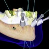 Computer Guided Treatment Planning and Surgery
Allesandro Pozzi, DDS
Robert Faulkner DDS
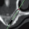
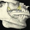 Angled Implants
Allesandro Pozzi, DDS
 Prosthodontic Procedures and Complications
Robert Faulkner, DDS