Transcript of “Ceramics in Fixed Prosthodontics – Considerations for use in Dental Practice”
1. Karl Lyons! Department of Oral Rehabilitation! Faculty of Dentistry Ceramics in Fixed Prosthodon2cs Considera2ons for use in Dental Prac2ce 1
2. Karl Lyons 2
3. Presentation Outline • Ceramics in dentistry • Types of ceramics • Clinical indications and uses • Case considerations • Preparation designs • Fitting and adjusting • Cementation • Longevity and causes of failure • Alternatives • Summary 3
4. Ceramics in Dentistry Why?
5. All Ceramic Crowns Zirconium Vs Leucite Reinforced Vs Metal-ceramic
6. Light Dynamics in Natural Teeth Tooth colours are produced by the colour of the dentine and pulp reflecting through the enamel layer which is influenced by the amount of demineralisation
7. Raptis et al. 2006 The Effect of Light Transmission Four crowns placed on tooth 11 Which two are PFM and which two are All-ceramic crowns?
8. In-Ceram Spinell IPS Empress PFM PFM (with porcelain shoulder) Note how light is blocked by the metal copings Raptis et al. 2006
9. Light Transmission
10. Charles Land • Invented dental porcelain in 1886 • Granted Patent 1887 • Platinum foil matrix Land CH (1903) Porcelain dental art: No II. Dental Cosmos 45:615-620 John McLean & TJ Hughes • Replaced metal reinforcement with Alumina (Al2O3) to develop first all- ceramic core The reinforcement of dental porcelain with ceramic oxides. (1965) BDJ 119(6):251-267 Evolution of Ceramic Crowns
11. Types of All-ceramic Monolithic crowns (1 layer) Bilayered crowns (2 layers – core & veneer) Eg: • Feldspathic –CAD/CAM blocks • Leucite reinforced – Empress I • Lithium Disilicate – emax Press or CAD • Zirconia (recently introduced) Eg: • Lithium Disilicate • Glass-infiltrated – In-Ceram Spinell • Alumina • Zirconia
12. Three main types of ceramics in dentistry 1 1. Predominantly glass • Veneering porcelains for PFM and all-ceramics • Most translucent – high aesthetics 2. Particle-filled glass (Glass-ceramics) i. High glass content • Lost wax system (Dicor) [no longer available] • Machinable feldspar-based ceramic (Vita Mark II blocks) • Heat-pressed Leucite reinforced (Empress I) (Kelly 2008)
13. Three main types of ceramics in dentistry 2 2. Particle-filled glass (Glass-ceramics) ii. Low glass content • Heat-pressed or CAD/CAM Lithium Disilicate (IPS emax) • Slip-cast or CAD/CAM Glass-infiltrated alumina (In-Ceram) 3. Polycrystalline (Ceramic oxides) i. Alumina Oxide (Procera Alumina) ii. Zirconia (3mol%Y-TZP) (Procera Zirconia)
14. Commercially Available Dental Ceramics
15. Flexural Strength of CeramicsFractureToughness[KIC(MPa.m1/2)] Flexural Strength (MPa)
16. Methods for Reinforcing Porcelain Metal-Oxides • Aluminium Oxide • Magnesium-Alumina Spinel • Zirconium Oxide Leucite Lithium Disilicate • Dispersion strengthening • Phase transformation toughening
17. VITABLOCS® Materials VITABLOCS Block Restoration Indication Mark II Inlays, onlays, anterior/posterior crown and veneer TriLuxe Anterior/posterior crown and veneer TriLuxe forte Anterior/posterior crown and veneer RealLife Anterior/posterior crown/ veneer for natural aesthetics
18. Structural Ceramics • Lithium disilicate • Excellent translucency and aesthetics • Inlays, onlays, veneers, crowns • In-Ceram Spinel • Excellent translucency and aesthetics • Anterior crown substructure
19. Structural Ceramics • In-Ceram Alumina • Good combination of strength and aesthetics • Substructure for anterior crowns and 3-unit bridges • In-Ceram Zirconia • Very good strength • Substructure for anterior and posterior crowns and 3-unit bridges
20. Structural Ceramics • In-Ceram AL • Very good strength • Substructure for anterior and 3-unit bridges and posterior crowns • In-Ceram YZ • Excellent strength • Substructure for anterior and posterior crowns and multi-unit bridges
21. Alumina and Zirconia • The increase in crystalline content in alumina and zirconia has: • Improved the mechanical properties allowing all-ceramic crowns and bridges • Is hard to machine and resistant to etching so resin bonding is a challenge
22. Zirconia
23. Zirconia
24. Note the difference in shrinkage between pre- sintered and sintered substructure. Zirconia Images compliments Vita Zahnfabrik
25. Procera
26. Overview of VITA In-Ceram® Ceramics
27. Vita Enamic
CAD/CAM Hybrid Ceramic
product description from Vita • For the first time, this innovative hybrid materials combines enormous strength with exceptional elasticity • As a result, the material is perfectly suited for crown restorations and moreover allows to achieve reduced wall thicknesses for minimally invasive restorations • Additionally, VITA ENAMIC excels by utmost reliability and precise and accurate milled restorations featuring high edge stability • This tooth-colored hybrid material also exhibits tooth- like material properties and produces highly esthetic results thanks to its excellent translucency
28. Ceramic network + Polymer network = Hybrid ceramic VITA ENAMIC: Dental hybrid ceramic! =! polymer-reinforced ceramic network Reinforced materials: (e.g. carbon, reinforced concrete)! = steel reinforced cement
29. Common Ceramic Core Materials Amorphous glass – Veneering porcelains Glass ceramics (reinforced by crystalline phases) • Leucite reinforced – Empress I • Lithium disilicate – Empress II • Magnesium aluminium oxide – In-Ceram Spinnell • Feldspathic Glass – Vita Mark II Blocks Glass infiltrated mixtures • In-Ceram alumina • In-Ceram zirconia Polycrystalline • Alumina – Procera • Zirconia – Lava, Everest, Cercon, Procera, Zeno, Ivoclar etc
30. 1. Amorphous glass – Vita Mark II 2. Crystalline glass ceramics (reinforced by crystalline phases) 1. Leucite reinforced – Empress I 2. Lithium disilicate – Empress II 3. Glass infiltrated mixture 1. Magnesium Aluminium Oxide – Spinell 2. InCeram alumina 3. InCeram zirconia 4. Polycrystalline 1. Alumina – Procera 2. Zirconia – Lava
31. All-Ceramic Material Type! Aesthetic Properties! Applications! Feldspathic Glass (predominantly glass) Intrinsically tooth coloured Anterior veneers & crowns Can be stained & glazed Crystalline Ceramics (particle-filled glass – high glass content) Intrinsically tooth coloured Anterior veneers & crowns Can be stained & glazed Glass Infiltrated Mixtures (particle-filled glass – low glass content) Core material Core can be pigmented Anterior and posterior crowns Are veneered with porcelain Polycrystalline Ceramics (no glass content) Core material Core can be pigmented Anterior and posterior crowns & bridges Are veneered with porcelain
32. 10! 8! 6! 4! 2! 0! 200! 400! 600! 800! 1000!0! FractureToughness(MPam1/2) Bending Strength (MPa) Filser et al., Quintessenz Zahntechnik 28, 1, 48-60 (2002) Empress 1 Dicor MGC In-Ceram Spinell In-Ceram Zirconia In-Ceram Alumina Zirconium oxide e.g. Zeno ZR IPS Empress 2 Glass Ceramics Crystalline Glass Ceramics Aluminium Oxide Zirconia Vita Mark II Aluminium Oxide Procera Anterior Crown Anterior Bridge All Restorations
33. Types of Ceramic Crowns Sintered porcelain • Feldspathic, Alumina or Leucite-reinforced Slip cast glass infused ceramic • In-Ceram – Alumina, Spinell or Zirconia Heat pressed ceramic – Leucite or Lithium disilicate • IPS Empress, IPS Empress 2 • Pentron OPC, Pentron OPC 3G • Finesse All-Ceramic Machined glass ceramic (eg CEREC 3, CEREC inLab) • Chair-side – VitaBlocs Mark II, ProCAD • Lab-milled – In-Ceram 2000, IPS e.max CAD, IPS Empress CAD Machined densely sintered ceramic – Alumina, Zirconia • Procera, Lava, Everest, e.max ZirCAD
34. Sintered Porcelain Crowns
35. • Feldspathic, Alumina & Leucite-Reinforced • Flexural Strengths • 55 – 75MPa (feldspar) • 90 – 120MPa (alumina) • 105 – 160MPa (leucite) • Clinical Survival Rates • Aluminous 73% over 5 years • Leucite • Anteriors 97% over 3 years • Posteriors 76% over 5 years Hankinson & Cappetta 1994; Etemadi & Smales 2006 Sintered Porcelain Crowns
36. Slip-Cast Glass Infused Ceramics
37. • In-Ceram: Alumina, Spinell & Zirconia • Flexural strengths • ~400MPa (Spinell) • ~500MPa (Alumina) • ~600MPa (Zirconia) • Clinical survival rates • In-Ceram Alumina & Spinell = 92% to 100% over 5 yrs • In-Ceram Zirconia = 3-unit FPD 95% over 3 yrs Wasserman et al. 2006 Slip-Cast Glass Infused Ceramics
38. Slip Casting Technique Images compliments Vita Zahnfabrik
39. Heat-Pressed Ceramic Crowns
40. IPS Empress, IPS Empress 2, Pentron OPC, Pentron OPC 3G, Finesse All-Ceramic • Flexural strengths – Leucite, Lithium disilicate • 140 – 200MPa (Leucite) • 350 – 450MPa (Li2O•2SiO2) • Clinical survival rates • Leucite 92% to 99% for anteriors over 3.5 years • Lithium disilicate 100% over 50 months (n=27) Chadwick 2004; Marquadt 2006 Heat-Pressed Ceramic Crowns
41. IPS Empress
42. Machined Glass-Ceramic Crowns
43. eg CEREC 3, CEREC inLab • Chair-side – VitaBlocs Mark II, ProCAD: • Lab-milled – In-Ceram 2000, IPS e.max CAD, IPS Empress CAD: • Industrially fabricated – stronger and more reliable • Clinical survival rates • Feldspathic-milled = 94% over 5 years (n=17, anterior) Bindl & Mormann 2004 Machined Glass-Ceramic Crowns
44. Machined Densely Sintered Crowns
45. Machined Densely Sintered Crowns • Alumina, Zirconia, Zircon • Procera, Lava, Everest, e.max ZirCAD • Flexural strengths • 600MPa (Alumina) • 700 – 820MPa (Zircon) • 850 – 1050MPa (Zirconia) • Clinical survival rates • Procera Alumina 92% at 10 years • Zirconia FPDs 100% of frameworks at 3 years Odman & Andersson 2001; Sailer et al. 2006
46. In-Ceram Alumina Empress2 Empress
47. Summary of Ceramics
48. Systematic Review
Goodacre et al 2003! Mean fracture rate for all-ceramic crowns increases as you move posteriorly Anteriors 3% Premolars 7% Molars 21% This review did not distinguish between: • fracture modes (core or veneer chipping) • or types of ceramic systems
49. Systematic Review
Pjetursson et al 2007 – All-ceramic vs PFM crowns 5yr survival rates: PFM 95.6% All-ceramic 93.3% 85% of all-ceramic crowns failures due to core fracture Chipping usually repairable Anteriors: All-ceramics = PFM Posteriors: Material dependent ü Alumina oxide 95% ü Reinforced glass ceramics (Empress) 94% ² In-Ceram 90% ² Glass-ceramics (Dicor) 85%
50. Systematic Review
Wang et al 2012 – All-ceramic single crowns 5 yr Fracture rate: (veneer + core) all systems Overall 7.7% Posteriors 10% Anterior teeth 4.4% Core fracture: Overall 7.2% Posteriors 9.5% Anteriors 3.9% Veneer chipping: Overall 3% Molars 3% Premolars 1.5% Canines 2.5% Incisors 2% No clear difference found Statistically significant Statistically significant
51. Systematic Review
Sailer et al 2007 – fixed partial dentures 5yr survival rates: Metal-ceramic FPDs 94.4% All-ceramic FPDs 88.6% Frequency of: core # veneer # Metal-ceramic FPDs 1.6% 2.9% All-ceramic FPDs 6.5% 13.6% Mainly Lithium disilicate and In-Ceram Rare in zirconia FPD Annual rate: Zirconia 1.98 – 12.2 Empress/emaxP 0.83 – 1.55 In-Ceram no chipping reported
52. Recommended Indications
Class 1 Ceramics • Aesthetic ceramic for coverage of a metal or ceramic subsurface and/or • Aesthetic ceramic for single-unit anterior, veneers, inlays, or onlays Example IPS Empress, IPS e.max Ceram (Ivoclar) Della Bona, 2009
53. Recommended Indications
Class 2 Ceramics • Aesthetic ceramic for adhesively cemented, single unit, anterior or posterior prostheses and/or • Adhesively cemented, substructure ceramic for single-unit anterior or posterior prostheses Example IPS Empress (Ivoclar), Cerec MkII (Vita) Della Bona, 2009
54. Recommended Indications
Class 3 Ceramics • Aesthetic ceramic for non-adhesively cemented, single-unit, anterior or posterior prostheses Example IPS e.max Press or CAD (Ivoclar) Della Bona, 2009
55. Recommended Indications
Class 4 Ceramics • Substructure ceramic for non-adhesively cemented, single-unit, anterior or posterior prostheses and/or • Substructure ceramic for three-unit prostheses not including molar restoration • Example IPS Empress 2, (Ivoclar), Cerec MkII (Vita) Della Bona, 2009
56. Factors that Influence Ceramics • Ceramics are susceptible to chemical corrosion and fatigue mechanisms • This reduces their lifetime • Unfavourable oral conditions include: • Chewing forces from 100-700 N • Moist environment at 37ºC • Small contact area; stresses generated 3.5-890 MPa Della Bona, 2009
57. Survival of Ceramics To improve mechanical behaviour of ceramics • Select the ceramic considering location • Consider substructure similar to metal for PFM • Minimise surface roughness • Rougher surfaces have more cracks so need fewer cycles of stress to fail • Chemical interaction between ceramic (crack tips) and environment (water) results in accelerated crack growth due to stress corrosion Della Bona, 2009
58. Considerations
in Fixed Prosthodontics
59. Considerations in Fixed Prosthodontics 3 The image cannot be displayed. Your computer may not have enough memory to open the image, or the image may have been corrupted. Restart your computer, and then open the file again. If the red x still appears, you may have to delete the image and then insert it again.
60. Crown Margin The image cannot be displayed. Your computer may not have enough memory to open the image, or the image may have been corrupted. Restart your computer, and then open the file again. If the red x still appears, you may have to delete the image and then insert it again.
61. Length of Edentulous Span 1 The image cannot be displayed. Your computer may not have enough memory to open the image, or the image may have been corrupted. Restart your computer, and then open the file again. If the red x still appears, you may have to delete the image and then insert it again.
62. Length of Edentulous Span 2 The image cannot be displayed. Your computer may not have enough memory to open the image, or the image may have been corrupted. Restart your computer, and then open the file again. If the red x still appears, you may have to delete the image and then insert it again.
63. Minimum occlusogingival and buccolingual connector dimensions as a function of position of the bridge connector and occlusal forces!
64. Ceramic Crown Form
65. The image cannot be displayed. Your computer may not have enough memory to open the image, or the image may have been corrupted. Restart your computer, and then open the file again. If the red x still appears, you may have to delete the image and then insert it again. Ceramic Crown Form The image cannot be displayed. Your computer may not have enough memory to open the image, or the image may have been corrupted. Restart your computer, and then open the file again. If the red x still appears, you may have to delete the image and then insert it again.
66. Minimum 9mm3 Tooth Preparation Summary
67. Bonding Ceramic Crowns • The crystalline content in alumina and zirconia is resistant to etching so bonding is a challenge • Silica coating systems (eg Rocatec and Cojet, 3M ESPE) creates a silica layer • High-speed surface impact of silica-modified alumina particles promotes resin bonding by: • Rough surface allowing micromechanical bonding to resin • Promotes a chemical bond between the silanated silica coated ceramic and the resin bond material
68. Bonding Strategies to Teeth and Restorative Materials
69. Commercially Available Dental Ceramics
70. Material properties: Strength Strength is probably the most advertised property in the dental literature.
71. 0 200 400 600 800 1000 1200 Zirconia Aluminium Oxide Lithium Disilicate – Hot Press Lithium Disilicate – CAD/CAM Magnesium Alliminium Oxide – Spinell Leucite Reinforced Feldspathic Glass Zirconia Aluminium Oxide Lithium Disilicate – Hot Press Lithium Disilicate – CAD/CAM Magnesium Alliminium Oxide – Spinell Leucite Reinforced Feldspathic Glass Series1 1100 620 470 350 400 175 80 Flexural Strength of All-Ceramic Materials in MPa Series1 Shinogaya et al, 2001, Clin Oral Invest 5:63-68
72. Failure Classifica2on Biological failures • Secondary caries Aesthetic failure • Restoration contours, value, hue, chroma Mechanical failures • Cracking • Chipping fracture (veneering porcelain fracture) • Bulk fracture (core substructure fracture) • Debonding of the prosthesis
73. Mechanical Failures • Cracking • Chipping of veneering porcelain • Cohesive chipping • Adhesive delamination • Bulk fracture (core substructure fracture) Transilumination Veneer porcelain chipping ! Delamination ! Core fracture!
74. Use Transillumination for Quality Control
75. Custom Fabrication • All-ceramic restorations are custom-fabricated increasing susceptibility to fabrication defects • Variety of techniques • Sintering • Heat-pressing • Slip-casting • CAD/CAM • Combined with staining or veneering step Each technique produces fabrication defects [from Janine Tiu]
76. Diamond grinding is a major source of failure-inducing flaws in dense ceramics. (Rice 2002) Zirconia CAD/CAM machining creates damage that is not fully healed by sintering process. (Kim et al 2010) Effects of Abrasive Grinding Thermal shock
77. Characteristic Trace Lines After CAD/CAM Machining! Lava! Procera Zr! Everest (KaVo)! DCS Zirkon!
78. Veneer Chipping in Zirconia-based Restorations Systematic reviews confirm chipping of veneering ceramic is the most frequent complication More common than with metal-ceramic or other all- ceramic restorations (Al-Amleh et al 2010, Hientz et al 2010, Schley et al 2010, Raigrodski et al 2012)
79. Al-Amleh et al 2010 “Clinical trials in zirconia: A systematic review Summary: • 17 clinical trials based on 3Y-TZP • Posterior FPD 13 studies • Single crowns 2 studies • Implant abutments 2 studies • 8 brands of zirconia • Longest trial 5yrs (only 2 studies) Chipping of veneering porcelain • Two of 15 studies did not report chipping • Was common for all brands • Incidence ranged from 0 – 54% • Not always noticed by patients – incidental finding • Also found at non-load bearing areas
80. 8 17
81. Difference between thermal conductivity between metal substructure and YZr substructure
82. Reasons for Zirconia Veneer Chipping High tensile residual stresses locked within the veneering porcelain (Swain 2009) Zirconia is a very poor thermal conductor: • Gold: 315 W/m-K • Alumina: 40 W/m-K • Zirconia: 2 W/m-K Substructure core design • Cap-like core do not support veneering porcelain • Suggested “PFM-style” cut back method [Tholey, Swain & Thiel 2011]
83. PFMs cool from the inside to the outside producing systematic compression bonding from the inside to the outside. YZr crowns cool from both the inside and the outside at a similar rate resulting in a compression layer in the outer veneer and YZr coping, and an inner zone of tension within the porcelain veneer. W/(m.K), of a gold coping = 315 W/(m.K), of a zirconia coping = 2 Thermal conductivity, W/(m.K), of porcelain = 1.4 Zone of tension
84. Loading Zirconia Crowns to Failure
fast cooled v slow cooled Procera Zirconia IPS e.max ZirPress Al-Amleh 2011
85. Fast Cooled Samples Common features: • Midline fissure crack • Cracking on mesial non-loaded side • Average 902 N
86. Fracture after 2 days Courtesy: Dr. L. Gruetter (University of Geneva)
87. Courtesy : Dr. L. Grütter (University of Geneva) Occlusal contact point responsible for shearing off veneering ceramic
88. 1. Clean (cotton pellet with alcohol), rinse & dry 2. Inject siloxane impression material (light body) 3. Cover the whole crown with silicon material Procera Alumina AllCeram (veneering ceramic failure after 4 years)
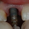
89. Zirconia Abutment Fracture Case description: 1. The zirconia implant abutment was screwed in tightly 2. Contact points M, D, were adjusted in situ 3. On the first bite for occlusal adjustment the crown fractured
90. Zirconia abutment CARES (Straumann) Zirconia Abutment Try-in Failure The first bite to check the occlusion created a stress concentration at the distal margin (white arrow). The crack path is marked by the red arrows and result in the crown splitting in half. origin Ø Fabrication defect
91. Cercon 6-unit bridge failure after 24 hours FPD Bulk (core) Fracture
92. Poor framework design • Not enough palatal clearance • Thin tip zirconia framework Both cause high stress concentrations Take home message: Always try-in zirconia frameworks before veneering
93. All zirconia copings were rejected
94. No substructure support Adequate substructure support allows for natural occlusal morphology
95. Procera Alumina All-Ceram (veneering ceramic failure after 4 years)
96. origi n Conclusion from the replica SEM analysis: The origin of the failure was located on the occlusal-palatal cusp (wear facette). The crack continued along the arrows downwards (interproximally) to the gingiva without reaching the margins. The veneering porcelain was unsufficiently supported by the alumina core
97. Guidelines for Restoring Chipped ! All-ceramic Restorations Grade 1: Fracture surface can be polished Grade 2: Fracture surface can be repaired with composite resin Grade 3: Severe fracture requires restoration replacement 1. Fracture extends into a functional area and repair is not feasible 2. Re-contouring will result in a significant unacceptable alteration of the anatomic form from the original anatomy 3. Re-contouring will significantly increase the risk of pulp trauma by the generation of heat 4. Repair with resin composite will result in unacceptable aesthetic result (Heintze & Rousson 2010, Anusavice 2012)
98. Success or Failure? • Is a chipped all-ceramic restoration a failed restoration? • Restoration success is defined as the demonstrated ability of a restoration to perform as expected • Acceptable surface quality • Anatomic contour • Function • Aesthetics (where applicable) • When should we repair or replace the entire restoration? • Restoration failure may be defined as any condition that leads to replacement of a prosthesis • Why do all-ceramic fracture? • How can we minimise this problem?
99. Origins of Fracture • Fabrication flaws of various shapes and sizes includes: • Pores Micro-cracks • Macro-cracks • Machining grooves • Air-abrasion surface defects • Grinding adjustments surface defects • Location of the defect under tensile stresses is important • Thermal residual stresses • Subcritical crack growth (SCCG) • In humid environment, cracks grow slowly but continuously Weakest link
100. Early v Late Fractures • Immediate failure or within a few hours or days of cementation is likely to originate from a major processing flaw (Schmitter et al 2009, Lohbauer et al 2010) • Failure after a few years is likely to involve subcritical crack growth and/or cyclic fatigue SCCG and/or cyclic fatigue
101. Most important factor affecting fracture rates: Position of restoration in the mouth Ferrario et al 2004 Greatest forces Molars > premolars > incisors (1/3-1/4 of molars load)
102. Surface area of incisal edge of anteriors and occlusal table of posteriors?
103. A F =σ Occlusal contacts on natural teeth are point contacts
104. Causes of Ceramic Substructure Failure • Fracture initiating in the connector area • Connector high stress area • Chipping of the veneering material • Residual stresses at the core-veneer interface • Differences in thermal conduction between the core and veneer • Thick veneer layer • Poor bonding between the core and veneer ceramic • Sliding occlusal contacts more damaging than axial contacts
105. Summary • Stronger ceramics are more opaque than aesthetic ceramics • Aesthetic restorations without much structural need – use single layer (monolithic) ceramics • High strength needed, less aesthetic ceramics veneers with tooth coloured porcelain • Any ceramic system suitable for veneers and anterior crowns • Only a few ceramics successful for restoring molars • Need to consider other clinical factors such as adequate preparation depth and cementation
106. Summary • No equivalent long-term data as for PFMs • ~75% at 15 years • Many ceramics >90% after 5 years • Reasonable evidence available for anterior 3- unit FPDs in lithium disilicate, In-Ceram Alumina and Zirconia • Posterior 3-unit FPDs only zirconia indicated • Chipping and fracture a problem • Higher success when ceramics bonded to teeth using resin cement rather than GIC • Use a silica coating system or primers for acid resistant ceramics such as zirconia
107. Ceramics in dentistry
108. Summary of Ceramics Recommendations Based on Peer Review Literature
109. Ceramics Aluminous Core Slip Cast – In-Ceram Hot Pressed – Empress Machined – Cerec , Procera
110. Ceramics
Slip Cast • Build-up with core particles and fired • High Strength • Highly Abrasive • Fair Marginal Fit – In-Ceram: crowns – In-Ceram Spinell: crowns (more translucent) – In-Ceram Zirconia: 3 unit FPD’s
111. In Ceram The image cannot be displayed. Your computer may not have enough memory to open the image, or the image may have been corrupted. Restart your computer, and then open the file again. If the red x still appears, you may have to delete the image and then insert it again. The image cannot be displayed. Your computer may not have enough memory to open the image, or the image may have been corrupted. Restart your computer, and then open the file again. If the red x still appears, you may have to delete the image and then insert it again. The image cannot be displayed. Your computer may not have enough memory to open the image, or the image may have been corrupted. Restart your computer, and then open the file again. If the red x still appears, you may have to delete the image and then insert it again.
112. What is Slip Casting?
113. Ceramics
Hot-Pressed • Medium to High Strength • Low to Medium Abrasive • Medium Translucency • Fair Marginal Fit – Empress & Finesse (leucite) ✦ veneers, inlays, onlays, crowns – Empress 2 (lithium disilicate) ✦ crowns, 3-unit FPD’s
114. What is Hot Pressed? Lost wax casting via pressed ceramic.
115. Ceramics
Machined (CAD/CAM) • Medium to Very High Strength • Low to Medium Abrasive • Fair Marginal Fit • Industrial CAD/CAM – Procera: crowns, FPDs • In-office CAD/CAM – Cerec: inlays – Cerec 2 & 3: inlays, onlays, crowns
116. IPS e.max KIc= 2.75 MPa√m KIc= 2.25 MPa√m LiO2 – SiO2 (lithium disilicate) S = 450 MPa S = 360 MPa T° = 850°CT° = 915-920°C
117. Images compliments Vita Zahnfabrik VITA In-Ceram® SPINELL
118. VITA In-Ceram® ALUMINA Images compliments Vita Zahnfabrik
119. VITA In-Ceram® ZIRCONIA Images compliments Vita Zahnfabrik
120. What is CAD/CAM ? Computer Aided Design / Computer Aided Manufacture CEREC® 3 inLab von Sirona inEos von Sirona
121. CAD/CAM ?
122. CAD/CAM
123. CAD/CAM
124. CAD/CAM
125. © Praxisfall Dr. Gunpei Koike (Japan) CAD/CAM
126. © Praxisfall Dr. Gunpei Koike (Japan) CAD/CAM
127. CAD/CAM
128. © Praxisfall Dr. Gunpei Koike (Japan) CAD/CAM
129. E4D HenrySchein Cerec Sirona Ivoclar Procera NobelBiocare CAD/CAM
130. All Ceramic Crowns • Indications – High cosmetic demand – Incisal edge reasonably intact – Favourable occlusion • Advantages – Cosmetics – Good tissue response – More conservative on labial
131. All Ceramic Crowns • Contraindications – High strength required – Insufficient tooth structure for support – Unfavourable occlusion • Disadvantages – Reduced strength – Not conservative – Brittle – Single crowns only
132. Types of crowns Full gold crown (FGC) Porcelain fused to metal (PFM) All ceramic
133. Affect of the Metal Coping The image cannot be displayed. Your computer may not have enough memory to open the image, or the image may have been corrupted. Restart your computer, and then open the file again. If the red x still appears, you may have to delete the image and then insert it again. The image cannot be displayed. Your computer may not have enough memory to open the image, or the image may have been corrupted. Restart your computer, and then open the file again. If the red x still appears, you may have to delete the image and then insert it again.
134. Ceramic Crown Form 1 The image cannot be displayed. Your computer may not have enough memory to open the image, or the image may have been corrupted. Restart your computer, and then open the file again. If the red x still appears, you may have to delete the image and then insert it again.
135. Occlusal Considerations The image cannot be displayed. Your computer may not have enough memory to open the image, or the image may have been corrupted. Restart your computer, and then open the file again. If the red x still appears, you may have to delete the image and then insert it again.
136. Ceramic Crown Form
137. Tooth Preparation 1 The image cannot be displayed. Your computer may not have enough memory to open the image, or the image may have been corrupted. Restart your computer, and then open the file again. If the red x still appears, you may have to delete the image and then insert it again. The image cannot be displayed. Your computer may not have enough memory to open the image, or the image may have been corrupted. Restart your computer, and then open the file again. If the red x still appears, you may have to delete the image and then insert it again.
138. Tooth Preparation 2 The image cannot be displayed. Your computer may not have enough memory to open the image, or the image may have been corrupted. Restart your computer, and then open the file again. If the red x still appears, you may have to delete the image and then insert it again. The image cannot be displayed. Your computer may not have enough memory to open the image, or the image may have been corrupted. Restart your computer, and then open the file again. If the red x still appears, you may have to delete the image and then insert it again. The image cannot be displayed. Your computer may not have enough memory to open the image, or the image may have been corrupted. Restart your computer, and then open the file again. If the red x still appears, you may have to delete the image and then insert it again. The image cannot be displayed. Your computer may not have enough memory to open the image, or the image may have been corrupted. Restart your computer, and then open the file again. If the red x still appears, you may have to delete the image and then insert it again.
139. All-ceramics and Metal-ceramics • The need to simulate in dental porcelains the light behaviour and appearance of the natural tooth • Create the illusion of nature within limited space constraints and light blocking effect of the metal substructure or ceramic substructure Yamamoto 1985 Yamamoto 1985
140. Think Cosmetics Bleaching Veneers Ceramic Crowns
141. Metal Ceramic Crowns ◆ Indications – Heavily restored / endodontically treated tooth – Cosmetics – When ceramic crowns not possible ◆ Advantages – Appearance compared with FGC – Retentive
142. Metal Ceramic Crowns ◆ Contraindications – Large pulp chamber – Intact buccal wall – More conservative restoration possible ◆ Disadvantages – Amount tissue removal – Occlusion difficult with glazed porcelain – Appearance compared with ceramic
143. Gold Crowns ◆ Indications – Heavily restored / endo treated tooth – Correct occlusal plane – Provide contours for RPD ◆ Advantages – Strong – Retentive
144. Gold Crowns ◆ Contraindications – < Maximum retention required – Cosmetics ◆ Disadvantages – Amount tissue removal – Metal display – Vitality testing
145. Dimensions of the Preparation The image cannot be displayed. Your computer may not have enough memory to open the image, or the image may have been corrupted. Restart your computer, and then open the file again. If the red x still appears, you may have to delete the image and then insert it again.
146. Light Transmission The image cannot be displayed. Your computer may not have enough memory to open the image, or the image may have been corrupted. Restart your computer, and then open the file again. If the red x still appears, you may have to delete the image and then insert it again.
147. Gold Crowns ◆ Indications – Heavily restored / endo treated tooth – Correct occlusal plane – Provide contours for RPD ◆ Advantages – Strong – Retentive
148. Gold Crowns ◆ Contraindications – < Maximum retention required – Cosmetics ◆ Disadvantages – Amount tissue removal – Metal display – Vitality testing
149. Preparation The image cannot be displayed. Your computer may not have enough memory to open the image, or the image may have been corrupted. Restart your computer, and then open the file again. If the red x still appears, you may have to delete the image and then insert it again.
150. The image cannot be displayed. Your computer may not have enough memory to open the image, or the image may have been corrupted. Restart your computer, and then open the file again. If the red x still appears, you may have to delete the image and then insert it again.
151. v Visit ffofr.org for hundreds of additional lectures on Complete Dentures, Fixed Prosthodontics Implant Dentistry, Removable Partial Dentures, Esthetic Dentistry and Maxillofacial Prosthetics. v The lectures are free. v Our objective is to create the best and most comprehensive online programs of instruction in Prosthodontics
New Lectures
 Angled Implants
Allesandro Pozzi, DDS
 Prosthodontic Procedures and Complications
Robert Faulkner, DDS
 Cement Retention vs Screw Retention
Robert Faulkner DDS, MS
 Single Tooth Defects in Posterior Quadrants
Robert Faulkner, DDS